Melanoma remains one of the most formidable challenges in oncology, a cancer notorious for its ability to spread early, mutate rapidly, and shrug off even the most sophisticated immunotherapies. Now, a team of researchers in China has uncovered a previously underappreciated molecular trick that the deadliest form of skin cancer uses to stay alive: a chemical tag on messenger RNA that keeps tumor cells protected from a form of programmed cell death known as ferroptosis. The study, published in Medical Oncology, identifies the RNA-binding protein YTHDF2 as a central player in melanoma progression and points to an unexpected candidate drug, the diabetes medication metformin, as a possible way to disarm it.
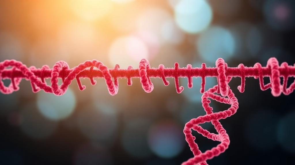
To understand why the finding matters, it helps to start with the chemistry. N6-methyladenosine, abbreviated m6A, is the most abundant internal modification found in eukaryotic messenger RNA. Rather than changing the genetic code itself, m6A acts like a sticky note attached to individual transcripts, influencing how long they survive, how efficiently they are translated into protein, and where they travel within the cell. The system relies on a cast of molecular actors: writers that deposit the methyl group, erasers that remove it, and readers such as YTHDF2 that interpret the mark and decide the fate of the tagged RNA. When YTHDF2 recognizes an m6A-modified transcript, it typically shepherds that mRNA toward degradation, effectively turning down the volume of the corresponding gene.
The research team, led by Da Gu, Huanmin Lou, Xiaojing Li, and colleagues at institutions including the First Affiliated Hospital of Anhui Medical University and Shandong First Medical University, began by mining public cancer databases. Their pan-cancer survey revealed that YTHDF2 is elevated across a range of tumor types, and in cutaneous melanoma specifically, its expression levels correlated positively with PD-L1, the molecular beacon that tumors use to suppress immune attack. Intriguingly, higher YTHDF2 also tracked with lower immune and stromal scores, suggesting that tumors rich in this RNA reader may build microenvironments that are less hospitable to infiltrating immune cells. That combination of immune evasion and aggressive growth made YTHDF2 a compelling suspect in the search for drivers of melanoma malignancy.
The next step was to ask what happens when YTHDF2 is removed. Using melanoma cell lines in the laboratory, the researchers knocked down the protein and observed a striking cascade of effects. Cell proliferation slowed markedly, and the population of Annexin V and propidium iodide positive cells, standard markers of dying cells, climbed. Crucially, when the team added a ferroptosis inhibitor to the cultures, the damage was largely rescued. That rescue experiment was the pivotal clue: it indicated that the cell death triggered by YTHDF2 loss was not ordinary apoptosis, but ferroptosis, an iron-dependent form of regulated cell death defined by the catastrophic oxidation of lipid membranes.
Ferroptosis has become one of the hottest topics in cancer biology precisely because tumor cells seem to fear it. The biochemical signature is unmistakable. In the YTHDF2-depleted melanoma cells, the researchers measured rising levels of intracellular ferrous iron, malondialdehyde, reactive oxygen species, and lipid peroxidation products, all hallmarks of membranes under oxidative assault. At the same time, glutathione, the cell’s principal antioxidant shield, dwindled. Every one of these changes was reversed when ferroptosis was pharmacologically blocked, confirming that the iron-fueled lipid destruction pathway was the mechanism at work. Experiments in animal models corroborated the same regulatory relationship in living tumors, lending physiological weight to the cell culture findings.
But how does an RNA-binding protein control a cell death pathway built from iron and lipids? The answer emerged from an integrated analysis combining RNA sequencing with RIP sequencing, a technique that maps which transcripts a protein physically binds. The data showed that YTHDF2 latches onto m6A marks on the messenger RNA of HMOX1, the gene encoding heme oxygenase 1, an enzyme with well-documented antioxidant and cytoprotective functions. By binding the methylated HMOX1 transcript, YTHDF2 destabilizes it, hastening its degradation and thereby suppressing the production of heme oxygenase 1. With less of this protective enzyme available, melanoma cells become more vulnerable to oxidative damage, yet the study’s overall picture shows that YTHDF2’s net effect in tumors is to promote malignant progression, positioning HMOX1 regulation as a key node in the network the protein controls.
The HMOX1 connection resonates with a growing body of literature. Heme oxygenase 1 has been implicated in ferroptosis regulation across diverse contexts, from liver cancer, where upregulating the enzyme synergizes with drug-induced ferroptotic stress, to retinal neovascularization and pulmonary hypertension, where YTHDF2 has separately been shown to suppress Hmox1-dependent antioxidant function in macrophages. The melanoma study now extends this theme into skin cancer, suggesting that the YTHDF2–HMOX1 axis is a recurring module that cells, whether diseased or malignant, use to tune their antioxidant capacity. In melanoma, the balance appears to tip in favor of tumor survival, consistent with YTHDF2’s established reputation as an oncogenic reader in cancers ranging from bladder carcinoma to diffuse large B-cell lymphoma.
Perhaps the most headline-grabbing element of the study is the repurposing angle. Metformin, a cheap, decades-old biguanide taken by millions of people with type 2 diabetes, has accumulated a long list of putative anticancer properties in the scientific literature. The researchers demonstrated that metformin inhibits YTHDF2 expression in melanoma cells, and in doing so promotes ferroptosis. The finding builds on the group’s earlier work, published in Cancer Genet, showing that metformin regulates ferroptosis in skin cutaneous melanoma through the ATF3/NRF2 axis. Together, the two studies sketch a coherent model in which the widely used drug pushes melanoma cells toward iron-mediated self-destruction by converging on the epitranscriptomic machinery that guards their antioxidant defenses. It also aligns with reports in breast cancer, where metformin induces ferroptosis by inhibiting the UFMylation of the cystine transporter SLC7A11, hinting that ferroptosis induction may be a unifying theme in the drug’s anticancer profile.
The therapeutic implications are tantalizing but must be tempered with appropriate caution. YTHDF2 has emerged as a target of growing interest in the field, with recent commentary in Trends in Cell Biology arguing that attacking the protein can reshape the epitranscriptome and overcome therapy resistance in tumors. A study in Science Immunology in 2024 detailed YTHDF2’s role in regulating immune evasion from within tumor cells, and the new melanoma data add a ferroptosis dimension to that immune story, given the observed correlation between YTHDF2 and PD-L1 expression. If blocking YTHDF2 simultaneously sensitizes tumor cells to ferroptosis and strips away immune camouflage, combination strategies that pair YTHDF2-directed approaches with checkpoint inhibitors become an obvious line of future investigation. Metformin, already proven safe in humans, could in principle be folded into such regimens more rapidly than a purpose-built YTHDF2 inhibitor, though clinical trials would be needed to establish any benefit in melanoma patients.
There are also broader lessons here about the m6A–ferroptosis interface, a frontier that a 2024 review in Molecular Cancer flagged as a promising avenue for advancing tumor immunotherapy. Other readers have been caught manipulating the same cell death pathway: in hepatocellular carcinoma, hypoxia was shown to block ferroptosis by suppressing the writer METTL14, which in turn unleashed YTHDF2-dependent silencing of SLC7A11. The melanoma study inverts the logic, with YTHDF2 acting on HMOX1 rather than SLC7A11, but the underlying principle is the same. Cancer cells exploit RNA modifications to fine-tune the expression of antioxidant genes, and each new target identified, whether a transporter, an enzyme like heme oxygenase 1, or a stress-response factor, adds a potential pressure point at which therapy could tip the balance toward tumor destruction. For a disease as stubborn as melanoma, that growing map of vulnerabilities is welcome news, and the humble RNA tag at the center of it may prove to be one of the most consequential sticky notes in modern cancer research.
Subject of Research: The role of the m6A reader YTHDF2 in regulating HMOX1-mediated ferroptosis and malignant progression in cutaneous melanoma
Article Title: YTHDF2 drives malignant progression in cutaneous melanoma through m⁶A-dependent HMOX1-mediated ferroptosis
Article References: Gu, D., Sun, Y., Sun, J., Kang, W., Lou, H., & Li, X. (2026). YTHDF2 drives malignant progression in cutaneous melanoma through m⁶A-dependent HMOX1-mediated ferroptosis. Medical Oncology, 43(11), Article 316. https://doi.org/10.1007/s12032-026-03427-y
Image Credits: AI Generated
DOI: 10.1007/s12032-026-03427-y
Keywords: melanoma, YTHDF2, m6A modification, ferroptosis, HMOX1, metformin, RNA methylation, PD-L1, lipid peroxidation, epitranscriptomics, skin cancer, cell death
Cite Scienmag News
Nathaniel Bowman. (October 10, 2026). Chemical Tag on RNA Helps Melanoma Dodge Ferroptosis, Study Finds. Scienmag. https://scienmag.com/chemical-tag-on-rna-helps-melanoma-dodge-ferroptosis-study-finds/
Nathaniel Bowman. "Chemical Tag on RNA Helps Melanoma Dodge Ferroptosis, Study Finds." Scienmag, 10 October 2026, https://scienmag.com/chemical-tag-on-rna-helps-melanoma-dodge-ferroptosis-study-finds/. Accessed 10 October 2026.
Nathaniel Bowman. "Chemical Tag on RNA Helps Melanoma Dodge Ferroptosis, Study Finds." Scienmag. October 10, 2026. https://scienmag.com/chemical-tag-on-rna-helps-melanoma-dodge-ferroptosis-study-finds/