For patients whose colorectal cancer has spread to the liver, one of the most agonizing questions is deceptively simple: will the targeted therapy actually work? Oncologists can currently offer only rough statistical averages, and the standard imaging yardstick, a rulebook known as RECIST 1.1, measures whether tumors shrink but says remarkably little about how long a patient will live. A new study published in BMC Cancer suggests that the answer may already be hiding inside routine MRI scans, encoded in texture patterns far too subtle for the human eye to grade.
A team of radiologists and researchers at Hubei Cancer Hospital and Wuhan Union Hospital, both affiliated with Tongji Medical College of Huazhong University of Science and Technology, developed and validated a hybrid prognostic model that fuses clinical data, conventional MRI features, and radiomics, a computational approach that converts medical images into thousands of quantifiable measurements. Their goal was ambitious: to predict overall survival in patients with colorectal cancer liver metastases, abbreviated CRCLM, before and during targeted therapy, using nothing more invasive than the scans patients already receive.
The study was retrospective and multicenter, drawing on 118 patients who underwent targeted therapy at the two tertiary hospitals. The investigators divided the cohort into a training set, an internal test set, and an external validation cohort, a design that matters enormously in the often-overhyped field of artificial intelligence in medicine. Many machine-learning models dazzle on the data they were built from and collapse when confronted with patients from a different hospital, different scanners, or different imaging protocols. By testing their model on an external cohort, the researchers deliberately exposed it to that harsher examination.
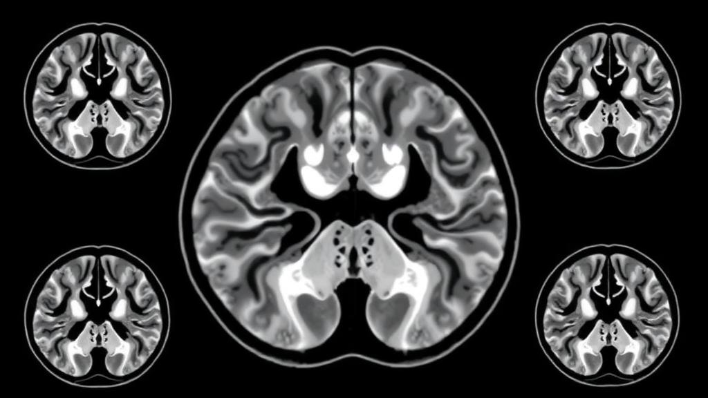
The technical heart of the study lies in how the team extracted information from the MRI images. Radiomics works by outlining a region of interest, in this case the tumor itself, and then computing mathematical descriptors of its appearance: statistical measures of how bright the pixels are, how uniform or chaotic their distribution, how coarse or fine the texture appears across different scales. These features capture biological properties that radiologists assess only subjectively, such as tumor heterogeneity, the internal diversity of a cancer that reflects genetically distinct subclones, variable blood supply, and patches of cell death. Heterogeneous tumors often behave more aggressively and respond less predictably to drugs, and radiomics offers a way to quantify that heterogeneity numerically rather than impressionistically.
What distinguishes this work is that the researchers did not stop at the tumor border. They built multiple models in parallel: one based on intratumoral features from inside the lesion, one based on peritumoral features from the tissue immediately surrounding it, a combined model, and so-called delta models that track how features change between scans over time. The peritumoral zone is biologically fascinating because it is where the tumor meets the immune system and the liver’s microenvironment, a battleground of invading cells, distorted vessels, and inflammatory signaling that often carries prognostic information invisible within the tumor core itself. Delta radiomics adds a temporal dimension, asking not just what the tumor looks like but how its texture evolves under the pressure of treatment, which may reveal whether a drug is genuinely reshaping the tumor’s biology.
After constructing these competing models, the team distilled them down and combined the best-performing radiomics score with key clinical and imaging variables. Two predictors emerged as independent drivers of overall survival: a clinical-imaging feature called TRTAV, tumor related to adjacent vein, which describes the spatial relationship between the metastasis and nearby hepatic veins, and the radiomics-derived Rad-score, a composite numerical signature of the tumor’s imaging texture. The vascular relationship is clinically meaningful because proximity to major veins is associated with more aggressive tumor biology and complicates surgical planning, while the Rad-score condenses the tumor’s internal and surrounding texture into a single risk-bearing number.
The resulting hybrid model outperformed every single-feature model, delivering robust accuracy in predicting survival at one, two, and three years across all cohorts, as measured by the area under the receiver operating characteristic curve, the standard statistic for gauging how well a model separates patients who experience an outcome from those who do not. Crucially, when the researchers split patients into low- and high-risk groups using the hybrid model, Kaplan–Meier analysis revealed statistically significant survival differences between the two strata in every comparison, with all p-values below 0.05. In plain terms, the model did not merely correlate with survival on average; it reliably sorted patients into groups whose survival curves visibly diverged.
Perhaps the most provocative claim in the paper is the comparison with RECIST 1.1, the response criteria that dominate oncology practice. RECIST classifies patients by whether their tumors shrink, stabilize, or grow, and it remains indispensable for regulatory decisions about drug approval. But tumor shrinkage is a crude proxy for benefit: some patients with modest radiographic change live far longer than others with dramatic responses. The hybrid model, the authors report, outperformed RECIST 1.1 in survival stratification, suggesting that quantitative imaging features capture prognostic information that simple size measurements discard. For a field searching for better ways to tell patients what to expect, that is a significant result.
The study also reflects the broader machinery of modern radiomics research. Feature selection tools such as minimum redundancy maximum relevance, known as mRMR, help prune thousands of candidate measurements down to a compact, non-overlapping set, guarding against the overfitting that plagues high-dimensional data. Interpretability techniques such as SHAP, SHapley Additive exPlanations, allow investigators to see which features pushed individual predictions up or down, addressing the black-box criticism that often dogs machine-learning medicine. The researchers also assessed intra- and inter-observer agreement, checking that different observers outlining the same tumors would extract consistent features, a mundane but essential safeguard for clinical translation.
Important caveats remain. The study enrolled 118 patients at two centers in a single country, and retrospective designs, however carefully controlled, cannot fully exclude selection effects. The model predicts overall survival under targeted therapy specifically, so its applicability to other treatments or cancer types is untested. External validation at additional institutions, ideally with prospective enrollment, will be needed before such a tool enters routine decision-making. The work received approval from both institutional review boards, with written informed consent from all patients, and was conducted under the Declaration of Helsinki and Good Clinical Practice guidelines.
Even with those limits, the study offers a glimpse of where cancer imaging is heading. Instead of asking whether a tumor shrank, clinicians may soon ask what a tumor’s texture, its vascular relationships, and its evolution under therapy reveal about the biology driving the disease. For patients with colorectal cancer that has reached the liver, a condition where treatment choices are consequential and prognoses vary widely, a non-invasive tool that turns a standard MRI into a personalized survival forecast could change conversations at the bedside. The scans were always there; the new work shows how much more they can be made to say.
Subject of Research: MRI-based delta radiomics for predicting overall survival to targeted therapy in colorectal cancer liver metastases
Article Title: Intratumoral and peritumoral delta radiomics of MRI predicts overall survival to targeted therapy in colorectal cancer with liver metastases
Article References: Xiao, R., Ai, S., Li, Y., Xiao, W., Liu, Z., Guo, X., & Liu, Y. (2026). Intratumoral and peritumoral delta radiomics of MRI predicts overall survival to targeted therapy in colorectal cancer with liver metastases. BMC Cancer. https://doi.org/10.1186/s12885-026-17014-x
Image Credits: AI Generated
DOI: 10.1186/s12885-026-17014-x
Keywords: colorectal cancer, liver metastases, radiomics, MRI, delta radiomics, targeted therapy, overall survival, prognostic model, RECIST 1.1, machine learning, tumor heterogeneity, TRTAV
Cite Scienmag News
Nathaniel Bowman. (October 10, 2026). MRI Radiomics Model Predicts Survival in Colorectal Cancer With Liver Metastases. Scienmag. https://scienmag.com/mri-radiomics-model-predicts-survival-in-colorectal-cancer-with-liver-metastases/
Nathaniel Bowman. "MRI Radiomics Model Predicts Survival in Colorectal Cancer With Liver Metastases." Scienmag, 10 October 2026, https://scienmag.com/mri-radiomics-model-predicts-survival-in-colorectal-cancer-with-liver-metastases/. Accessed 10 October 2026.
Nathaniel Bowman. "MRI Radiomics Model Predicts Survival in Colorectal Cancer With Liver Metastases." Scienmag. October 10, 2026. https://scienmag.com/mri-radiomics-model-predicts-survival-in-colorectal-cancer-with-liver-metastases/