


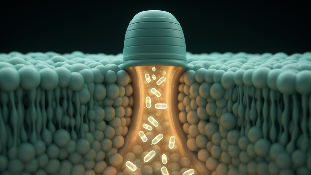
In a striking twist of immunological irony, scientists have discovered that the very process a cell uses to destroy itself in flames of inflammation may also open the door to its own rescue. Pyroptosis, the explosive form of inflammatory cell death, begins when enzymes called inflammatory caspases punch thousands of microscopic holes in the cell membrane. New research published in Nature by Groborz and colleagues, and highlighted in Cell Research by Órlaith C. Henry and Luke A. J. O’Neill of Trinity College Dublin, shows that these holes, formed by the protein gasdermin D, can serve as entry gates for precisely designed drugs that shut the deadly process down. The finding reads like a molecular Trojan horse story: the cell’s own self-destruction machinery ushers the enemy inside, only for that enemy to disarm the attack from within.
Pyroptosis is not an ordinary death. It is a programmed, highly inflammatory form of cell suicide that serves as an alarm system for the immune system. It is triggered when pathogen-associated or danger-associated molecular patterns are detected in the cytosol, the fluid interior of the cell. Upon sensing these threats, caspase-1 in humans and mice, along with caspase-4, caspase-5 in humans and caspase-11 in mice, cleaves a protein called gasdermin D, or GSDMD. This cleavage releases the N-terminal fragment of GSDMD, which migrates to the cell membrane and oligomerizes into pores. These pores disrupt the osmotic balance of the cell, allowing water to rush in, causing the cell to swell, burst and spill its inflammatory contents, including the potent cytokine interleukin-1β, into the surrounding tissue.
While this fiery demise is useful for containing infection, overactive pyroptosis is a driver of disease. Excessive pyroptotic cell death has been implicated in atherosclerosis, arthritis, Alzheimer’s disease, HIV and various types of cancer. This has made the inflammatory caspases attractive drug targets, and caspase inhibitors have captured the attention of scientists and clinicians alike. Yet developing safe and clinically effective caspase inhibitors has proven remarkably difficult. The central problem is selectivity. Caspases are a family of proteases with overlapping substrate preferences, which makes it difficult to inhibit one member without affecting others. Many existing inhibitors block caspases involved in both pyroptosis and apoptosis, the quiet, non-inflammatory form of cell death that is essential for development and immunity. Worse, inhibiting caspase-8 can sensitize cells to necroptosis, another damaging form of cell death, potentially trading one pathology for another.
To escape this trap, Groborz and colleagues took a rational, chemistry-driven approach. They exploited the known substrate preferences of individual caspases to synthesize a library of roughly one hundred tetrapeptides, each attached to an acyloxymethyl ketone, or AOMK, group. These reactive chemical groups allow the tetrapeptides to bind covalently and irreversibly to the active sites of their target caspases, ensuring durable inhibition. From this library, several candidates emerged that suppressed cell death driven by caspase-1, caspase-4 and caspase-11 while leaving staurosporine-induced apoptosis untouched. One of the most potent, named KGR-3, stood out as a strong inhibitor of caspase-1 and caspase-4.
The detailed characterization of KGR-3 revealed just how precisely it worked. The compound blocked pyroptosis by preventing the cleavage of GSDMD, thereby stopping pore formation at its source. It also attenuated the release of interleukin-1β and lactate dehydrogenase, a standard marker of membrane rupture, in response to activation of the NLRP3 and NLRP1 inflammasomes, the multiprotein complexes that activate caspase-1. Crucially, KGR-3 did not interfere with hallmarks of apoptosis such as caspase-1-induced blebbing or caspase-3 cleavage, nor did it affect necrosis triggered by listeriolysin O, a bacterial toxin. Cells treated with KGR-3 not only avoided death; their swelling and lysis were suppressed and they continued to grow and divide. The inhibitor appeared to grant cells a genuine reprieve rather than merely delaying the inevitable.
Yet a puzzling observation threatened to undermine the whole story. KGR-3 and its chemical siblings showed limited permeability in healthy cells, meaning they should not have been able to reach the intracellular caspases they so effectively inhibited. This paradox led the authors to a bold hypothesis: pyroptosis itself facilitates inhibitor uptake. Like a Trojan horse being wheeled through the gates of a besieged city, the caspase inhibitors were exploiting the very pores formed during pyroptosis to slip inside and reach the caspases driving the process. To test this idea, the researchers used propidium iodide and YOYO-1, two small fluorescent molecules that normally cannot cross intact membranes. Both entered cells in a GSDMD-dependent manner, providing strong evidence that gasdermin pores admit small molecules from the outside.
The story became even more sophisticated when the researchers examined what happened to the membrane damage inflicted along the way. In KGR-3-treated cells, GSDMD-induced membrane injury was repaired by the endosomal sorting complexes required for transport, known as the ESCRT machinery. This repair system is a conserved cellular mechanism that seals small membrane wounds. When the researchers expressed dominant-negative versions of the ESCRT proteins VPS4A and CHMP3, KGR-3 lost its protective effect, and pyroptosis proceeded. This finding challenges the simple binary view of pyroptosis as an irreversible commitment to death. Instead, it suggests a pharmacologically accessible window between the initiation of pyroptosis and final lysis, a period during which GSDMD pores disrupt the membrane enough to admit the drug, but not so extensively that repair by ESCRT becomes impossible. Recent research has independently highlighted the reversibility of GSDMD pore formation and shown that ESCRT can limit pyroptosis and cytokine secretion after inflammasome activation, lending further support to this model.
There is also an elegant pharmacological lesson hidden in the poor permeability of these compounds. In traditional drug development, limited cell permeability is considered a weakness. Here, it becomes a feature. Because the inhibitors can only enter cells whose membranes have already been breached by gasdermin pores, they are restricted to unhealthy cells, those actively undergoing pyroptosis, and are excluded from healthy neighbors. This built-in targeting mechanism could limit the off-target effects that have plagued broader caspase inhibitors, offering a new design principle: drugs need not always be membrane permeable if the disease state itself creates the doorway. The study thus overturns the conventional assumption that any drug aimed at an intracellular target must be able to cross an intact plasma membrane.
Translating the concept into animals required further chemical refinement. KGR-3 itself showed limited efficacy in vivo, but another tetrapeptide from the library, KGR-53, proved more promising. It was selected for animal testing because of its potent inhibition of caspase-11, a key mediator of endotoxic shock induced by lipopolysaccharide in mice. Because negatively charged compounds are transported inefficiently through GSDMD pores, the team designed a derivative called KGR-53P that lacks a negative charge. This optimized compound inhibited GSDMD cleavage and blocked the caspase-1- and caspase-11-dependent secretion of interleukin-1β and interleukin-18, while leaving both intrinsic and extrinsic apoptosis intact. In a mouse model of endotoxic shock, a condition driven by runaway caspase-11 activity, this selectivity could prove decisive.
The work is not without caveats, as Henry and O’Neill note in their highlight. First, the entry of KGR-3 through GSDMD pores has not been directly visualized. Although the GSDMD-dependent uptake of propidium iodide and YOYO-1 strongly implies that the pores serve as entry routes for small molecules, attempts to image KGR-3 itself failed because fluorescent tagging compromised the compound’s potency, permeability or toxicity. Second, KGR-53 showed evidence of liver toxicity in vivo, and it remains unclear whether this toxicity stems from the AOMK chemistry, an off-target effect or an entirely different mechanism. These open questions will shape the next phase of research. Even so, the conceptual advance is considerable. If the window between pyroptosis initiation and lysis can be reliably exploited, a new class of anti-inflammatory drugs could emerge that intervene only where inflammation is already burning, leaving healthy tissue untouched. The gates that pyroptosis opens may, in the end, become the route by which medicine tames it.
Subject of Research: Selective inflammatory caspase inhibitors that enter cells through gasdermin D pores to inhibit pyroptosis
Article Title: A Trojan horse story: does pyroptosis open the gates to its own inhibition?
Article References: Henry, Ó. C., & O’Neill, L. A. J. (2026). A Trojan horse story: does pyroptosis open the gates to its own inhibition?. Cell Research. https://doi.org/10.1038/s41422-026-01302-z
Image Credits: AI Generated
DOI: 10.1038/s41422-026-01302-z
Keywords: pyroptosis, gasdermin D, inflammatory caspases, caspase-1, caspase-11, GSDMD, ESCRT, inflammasome, cell death, innate immunity, drug design, inflammation
Cite Scienmag News
William Thompson. (October 8, 2026). Trojan Horse Inhibitors Slip Through Gasdermin Pores to Shut Down Pyroptosis. Scienmag. https://scienmag.com/trojan-horse-inhibitors-slip-through-gasdermin-pores-to-shut-down-pyroptosis/
William Thompson. "Trojan Horse Inhibitors Slip Through Gasdermin Pores to Shut Down Pyroptosis." Scienmag, 8 October 2026, https://scienmag.com/trojan-horse-inhibitors-slip-through-gasdermin-pores-to-shut-down-pyroptosis/. Accessed 8 October 2026.
William Thompson. "Trojan Horse Inhibitors Slip Through Gasdermin Pores to Shut Down Pyroptosis." Scienmag. October 8, 2026. https://scienmag.com/trojan-horse-inhibitors-slip-through-gasdermin-pores-to-shut-down-pyroptosis/


