


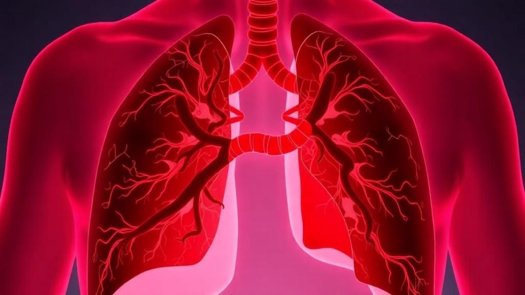
Pulmonary arterial hypertension, a rare and relentlessly progressive disease in which the blood vessels of the lungs narrow and stiffen until the right side of the heart fails, has long been treated as a disorder of vascular tone. Drugs that dilate those vessels through the prostacyclin, nitric oxide, and endothelin pathways have extended and improved lives, yet they do little to reverse the cellular remodeling that keeps the disease moving forward. A new narrative review published in Molecular Biology Reports argues that the field has been overlooking a molecular culprit that sits upstream of much of that remodeling: oxidized mitochondrial DNA, a fragment of the cell’s own genetic material that, once damaged and released, behaves less like debris and more like a persistent alarm signal that keeps the vasculature in a state of self-perpetuating injury.
The review, authored by a team at the All India Institute of Medical Sciences in New Delhi and collaborators at Delhi Pharmaceutical Sciences and Research University, synthesizes converging experimental and clinical evidence that pulmonary arterial hypertension is fundamentally a mitochondria-centered disease. In the pulmonary arterial endothelial and smooth muscle cells of patients, energy metabolism shifts early and dramatically, redox balance collapses, and the organelles that should be quietly producing ATP instead begin leaking danger signals into the cytosol. The authors contend that oxidized mitochondrial DNA is not merely a passive byproduct of this mitochondrial injury but an active mechanistic driver that sustains the hyperproliferation and apoptosis resistance characteristic of diseased pulmonary vascular cells.
To understand why this molecule is so potent, it helps to consider its double identity. Mitochondrial DNA descends from bacterial ancestors, and the innate immune system has never forgotten that heritage. It retains receptors, including Toll-like receptor 9 and the cGAS-STING pathway, that treat cytosolic or extracellular DNA as evidence of infection. When mitochondrial DNA is chemically oxidized, its inflammatory potency increases further, and it becomes a ligand that activates the NLRP3 inflammasome, driving caspase-1 to cleave pro-IL-1β into its mature form and gasdermin D to form pores in the cell membrane. In other words, a molecule that normally stays locked inside mitochondria can, once oxidized and escaped, switch on the same inflammatory machinery that responds to pathogens.
The review describes how the disease environment loads the dice toward exactly that outcome. Chronic hypoxia, inflammatory cytokines, altered shear stress in the vessel wall, and genetic vulnerability, including mutations in BMPR2, converge to amplify reactive oxygen species production inside mitochondria. That oxidative burst damages the mitochondrial genome directly, and oxidative lesions make the DNA more bioactive and more persistent once it escapes. At the same time, the systems that should clear damaged DNA and damaged organelles, the nucleases and the autophagy-lysosome machinery, become overloaded, allowing oxidized mitochondrial DNA to accumulate rather than be quietly disposed of. The result is a feed-forward loop: oxidative stress produces oxidized mitochondrial DNA, which drives inflammatory activation, and the cytokine and interferon signaling generated by that inflammation further weakens antioxidant defenses and mitochondrial respiration, generating still more reactive oxygen species.
The escape routes for mitochondrial DNA are themselves a subject of mechanistic interest. The review catalogs several pathways by which the molecule exits its home organelle, including leakage through the mitochondrial permeability transition pore and voltage-dependent anion channels, export inside mitochondria-derived vesicles, and release through sublethal permeabilization of the outer mitochondrial membrane. Each route matters because each represents a potential point of therapeutic intervention. If the pores and channels through which oxidized DNA escapes can be stabilized or blocked, the inflammatory cascade that follows might be blunted at its source rather than suppressed downstream with broad immunosuppression.
Perhaps the most conceptually significant section of the review concerns the autophagy-mitophagy axis, the cellular quality-control system that normally identifies damaged mitochondria, tags them with ubiquitin through the PINK1-Parkin pathway, and digests them in lysosomes. In pulmonary arterial hypertension, this system becomes dysregulated in a particularly insidious way: autophagic flux is reduced and signaling is skewed, so injured mitochondria are neither efficiently cleared nor allowed to die. Instead, they persist as signal emitters, continuously releasing oxidized mitochondrial DNA and reactive oxygen species while the cell remains alive and, worse, continues to proliferate. This reframes mitophagy failure not as a secondary consequence of disease but as a mechanism that actively sustains it, and it suggests that restoring mitophagic flux could interrupt the cycle at a fundamental level.
Calcium signaling forms a third arm of the pathological circuit. The review details how oxidized mitochondrial DNA and the stress it generates remodel endoplasmic reticulum-mitochondria contact sites, known as MAMs, which coordinate calcium transfer between the two organelles. In diseased pulmonary vascular cells, L-type calcium channels are upregulated, sarcoplasmic reticulum handling through channels such as RyR2 is altered, and store-operated calcium entry is enhanced, all of which raise cytosolic calcium concentrations. Elevated calcium drives contraction, proliferation, and further mitochondrial calcium overload, which in turn sensitizes the permeability transition pore and promotes more DNA release. Calcium dysregulation, mitochondrial damage, and inflammation thus reinforce one another in a loop that the review’s authors place at the center of their disease model.
The clinical implications are twofold. First, oxidized mitochondrial DNA could serve as a biomarker. Circulating levels of the molecule have been correlated with disease severity in cross-sectional human sampling, and related work in other cardiovascular contexts, including a longitudinal cohort in psoriasis patients, has linked circulating oxidized mitochondrial DNA broadly to cardiovascular disease. A readily measurable blood marker that reflects the mitochondrial and inflammatory state of the pulmonary vasculature would be a genuine advance in a disease currently monitored largely through hemodynamic measurements. Second, the pathways the review maps, from the NLRP3 inflammasome and cGAS-STING signaling to calcium channels and mitophagy regulators, offer a menu of therapeutic targets that address the remodeling process itself rather than merely relaxing vessels. Early clinical signals, such as pilot studies of interleukin-1 blockade in pulmonary arterial hypertension, hint that targeting inflammation may be feasible, and mitochondria-targeted antioxidants are being explored across cardiopulmonary disease.
The authors are careful, however, to flag the limits of the current evidence base. Most of the mechanistic data linking oxidized mitochondrial DNA to autophagy-mitophagy dysregulation come from cross-sectional human samples and from disease models that are not pulmonary arterial hypertension itself, and the correlations between circulating oxidized mitochondrial DNA and disease severity have not yet been tested in prospective, longitudinal PAH cohorts. Until such validation is completed, they argue, the biomarker potential of oxidized mitochondrial DNA should be treated as hypothesis-generating rather than established. That caution is appropriate for a review, but it does not diminish the framework’s appeal. By positioning a single molecule at the junction of mitochondrial stress, innate immunity, calcium homeostasis, and quality-control failure, the review offers researchers a unifying model of a disease that has long been described in fragments, and it identifies a concrete set of experiments, and eventually trials, that could determine whether the cell’s own damaged genome can be transformed from a driver of pulmonary arterial hypertension into its most informative biomarker.
Subject of Research: The role of oxidized mitochondrial DNA in autophagy-mitophagy dysregulation and pulmonary vascular remodeling in pulmonary arterial hypertension
Article Title: Oxidized mitochondrial DNA as a potential biomarker and mechanistic driver of autophagy–mitophagy dysregulation in pulmonary arterial hypertension: a narrative review
Article References: Dubey, N., Singh, S., Panchbhai, P., Chaturvedi, G., Parakh, N., Yadav, R. K., Kumar, S., & Yadav, H. N. (2026). Oxidized mitochondrial DNA as a potential biomarker and mechanistic driver of autophagy–mitophagy dysregulation in pulmonary arterial hypertension: a narrative review. Molecular Biology Reports, 53(1), Article 1716. https://doi.org/10.1007/s11033-026-12895-4
Image Credits: AI Generated
DOI: 10.1007/s11033-026-12895-4
Keywords: pulmonary arterial hypertension, oxidized mitochondrial DNA, mitochondrial dysfunction, autophagy, mitophagy, NLRP3 inflammasome, cGAS-STING, calcium signaling, reactive oxygen species, biomarker, pulmonary vascular remodeling, innate immunity
Cite Scienmag News
Drew Townsend. (October 12, 2026). Damaged Mitochondrial DNA Emerges as a Central Driver of Pulmonary Arterial Hypertension. Scienmag. https://scienmag.com/damaged-mitochondrial-dna-emerges-as-a-central-driver-of-pulmonary-arterial-hypertension/
Drew Townsend. "Damaged Mitochondrial DNA Emerges as a Central Driver of Pulmonary Arterial Hypertension." Scienmag, 12 October 2026, https://scienmag.com/damaged-mitochondrial-dna-emerges-as-a-central-driver-of-pulmonary-arterial-hypertension/. Accessed 12 October 2026.
Drew Townsend. "Damaged Mitochondrial DNA Emerges as a Central Driver of Pulmonary Arterial Hypertension." Scienmag. October 12, 2026. https://scienmag.com/damaged-mitochondrial-dna-emerges-as-a-central-driver-of-pulmonary-arterial-hypertension/


