


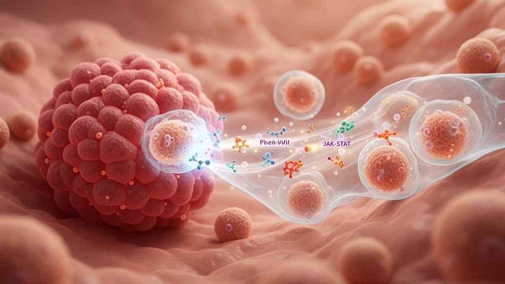
A 2026 review identifies aberrant JAK-STAT signaling as a major mechanism shaping the immunosuppressive microenvironment of breast cancer, particularly triple-negative breast cancer, while cautioning that drugs against the pathway have not yet produced convincing clinical responses in existing trials. The analysis describes how STAT3 and STAT5 can raise PD-L1 expression, expand myeloid-derived suppressor cells and regulatory T cells, weaken CD8+ T-cell and natural killer-cell activity, and support tumor growth, invasion, stemness and immune escape. It also argues that combination with immune checkpoint inhibitors remains a promising but unproven strategy requiring more selective agents, better patient selection and larger clinical validation.
JAK-STAT is a signaling cascade used by cytokines, growth factors and hormones to transmit information from cell-surface receptors into the nucleus. Binding of a cytokine to its cognate receptor brings receptor-associated Janus kinases into proximity, allowing them to activate one another by trans-phosphorylation. The kinases then phosphorylate signal transducers and activators of transcription, or STAT proteins, which form dimers, enter the nucleus and bind specific DNA sequences in target-gene promoters.
The family contains four Janus kinases—JAK1, JAK2, JAK3 and TYK2—and seven STAT proteins. Negative regulation involves SOCS/CIS proteins, protein inhibitors of activated STAT and protein tyrosine phosphatases, whose loss can allow persistent signaling and disrupted immune balance. Not all STAT proteins have the same immunological effect.
STAT1, a downstream mediator of type I and type II interferon signaling, promotes MHC class I expression, release of the T-cell chemokines CXCL9 and CXCL10, and cytotoxic activity of T and natural killer cells. STAT3 and STAT5 are more strongly associated with immunosuppression in breast cancer. STAT3 can directly promote transcription of PD-L1, the gene also known as CD274, and support the recruitment and differentiation of myeloid-derived suppressor cells.
STAT5 responds to cytokines including IL-2, IL-7 and IL-15, drives FOXP3 expression and supports the differentiation, survival and suppressive function of regulatory T cells. The review also notes a complication: STAT1 and STAT3 can form heterodimers that jointly bind the PD-L1 promoter, so blocking either protein alone reduces its expression only partially. The patterns of abnormal activation differ among breast cancer subtypes.
In triple-negative disease, interleukin-6 acting through IL-6R and JAK1/JAK2 can activate STAT3, increase PD-L1 and support cancer stem-cell properties, invasion and metastatic capacity. This signaling also recruits myeloid-derived suppressor cells and creates a self-amplifying immunosuppressive loop. By contrast, HER2-positive tumors can activate STAT3 through SRC kinase and JAK2, forming a survival axis that may contribute to resistance to trastuzumab.
In luminal breast cancer, prolactin-driven JAK2-STAT5a signaling can intersect with estrogen-receptor pathways, whereas persistent STAT3 activation is associated with endocrine resistance and a shift toward a more immunosuppressive tumor state. These differences mean that pathway activity cannot be treated as a uniform feature across breast cancer. Several mechanisms allow JAK-STAT signaling to influence malignant progression beyond immune regulation.
STAT3 and STAT5 can increase genes such as cyclin D1 and MYC while supporting anti-apoptotic proteins including BCL2 and BCL-XL. STAT3 can induce epithelial-mesenchymal transition and raise matrix metalloproteinases MMP-2 and MMP-9, while also activating stemness genes such as NANOG and OCT4 through an IL-6 autocrine loop. It can promote angiogenesis through vascular endothelial growth factor, and endothelial JAK-STAT signaling can regulate vascular barrier function, endothelial migration and the trafficking of immune cells.
In breast cancer models and patient samples, pharmacologic inhibition or RNA interference against STAT3 has been associated with reduced PD-L1 expression; inhibiting STAT3 has also increased antitumor macrophage phenotypes and natural killer-cell activation in experimental systems. Clinically, the translation of these findings has been more difficult than the preclinical biology might suggest. In a phase II study of ruxolitinib monotherapy in metastatic triple-negative breast cancer, none of 21 evaluable patients achieved an objective response and median progression-free survival was 1.2 months.
Paired biopsies nevertheless showed a significant reduction in phosphorylated STAT3-positive cells, indicating that the drug engaged its intended target. The review attributes this disconnect to compensatory survival signaling through pathways such as PI3K/AKT and MAPK, as well as to suppression of JAK1-STAT1 interferon signaling needed for antigen presentation and CD8+ T-cell activation. Ruxolitinib treatment has also been associated with reduced intratumoral CD8+ T cells in the reviewed evidence, underscoring why broad pathway inhibition may suppress protective immunity as well as tumor-promoting signals.
More selective STAT-directed treatments are being developed in response to these limitations. Direct STAT3 inhibition remains difficult because the protein’s SH2 domain must recognize phosphorylated receptor motifs, a function that is central to both recruitment and dimer formation. Peptide-based inhibitors can bind strongly but may be degraded rapidly and cross cell membranes poorly; non-peptide molecules generally face a trade-off among affinity, permeability and stability.
The review reports that no SH2-domain inhibitor has received US Food and Drug Administration approval. Proteolysis-targeting chimeras offer another route by recruiting an E3 ubiquitin ligase to degrade STAT3. Preclinical compounds have shown selective degradation, and KT-333, a STAT3 degrader, has entered phase I trials in hematologic malignancies and solid tumors, although its efficacy and safety specifically in breast cancer remain unknown.
Combination strategies have likewise produced mixed results. A phase I/II trial of ruxolitinib plus trastuzumab in patients with resistant HER2-positive disease reported median progression-free survival of 8.3 weeks, below expectations. At the Mayo Clinic, a phase I trial combined ruxolitinib with pembrolizumab in 12 patients with chemotherapy-refractory metastatic triple-negative breast cancer.
No patient had an objective response, median time to progression was three months, and two patients at lower dose levels had stable disease for six months. Adverse effects included elevated transaminases and anemia or leukopenia in all 12 participants, fatigue in nine and grade 3 or higher events in five. A separate phase II study cited in the review found that ruxolitinib suppressed phosphorylated STAT3 but also reduced granzyme-B-positive T cells, suggesting that immune remodeling did not necessarily translate into a benefit for checkpoint blockade.
Patient selection may be essential because triple-negative breast cancer is molecularly heterogeneous. The review identifies immune-infiltrated disease as more likely to benefit from checkpoint blockade than immunosuppressive or mesenchymal subtypes, but no ideal predictive biomarker is available. Phosphorylated STAT1 positivity, 9p24.1 amplification involving JAK2 and PD-L1, immune-cell signatures such as a CTL-NK score, and circulating tumor-cell PD-L1 are candidate approaches rather than validated selectors.
PD-L1 immunohistochemistry alone has limited ability to distinguish beneficiaries, and STAT3 activation is prognostic rather than proven predictive of treatment response. Researchers also need to account for differences among primary and metastatic lesions and for changes over time. The review concludes that selective inhibition of immunosuppressive STAT3 or STAT5 signaling, alongside careful preservation of STAT1-mediated surveillance, must be tested in complementary models—including syngeneic systems, humanized mice, patient-derived xenografts and organoids—before combination regimens can be considered clinically established.
Subject of Research: Review of JAK-STAT signaling as a target for remodeling breast cancer immunity and developing combination immunotherapy.
Article Title: JAK-STAT signaling: a key mechanism of immune microenvironment remodeling in breast cancer and novel strategies for targeted therapy
Article References: Qiao, J., Sun, T., Wang, G., Yan, Y., & Tong, W. (2026). JAK-STAT signaling: a key mechanism of immune microenvironment remodeling in breast cancer and novel strategies for targeted therapy. Frontiers in Oncology, 16, Article 1947188. https://doi.org/10.3389/fonc.2026.1947188
Image Credits: AI Generated
DOI: 10.3389/fonc.2026.1947188
Keywords: breast cancer, JAK-STAT signaling, tumor immune microenvironment, STAT3, STAT5, immune checkpoint inhibitors, triple-negative breast cancer, PD-L1, cancer immunotherapy, myeloid-derived suppressor cells, JAK-STAT, signaling
Cite Scienmag News
Nathaniel Bowman. (October 21, 2026). Research report: JAK-STAT signaling: a key mechanism of immune microenvironment remodeling in breast cancer and novel strategies for targeted therapy. Scienmag. https://scienmag.com/research-report-jak-stat-signaling-a-key-mechanism-of-immune-microenvironment-remodeling-in-breast-cancer-and-novel-strategies-for-targeted-therapy/
Nathaniel Bowman. "Research report: JAK-STAT signaling: a key mechanism of immune microenvironment remodeling in breast cancer and novel strategies for targeted therapy." Scienmag, 21 October 2026, https://scienmag.com/research-report-jak-stat-signaling-a-key-mechanism-of-immune-microenvironment-remodeling-in-breast-cancer-and-novel-strategies-for-targeted-therapy/. Accessed 21 October 2026.
Nathaniel Bowman. "Research report: JAK-STAT signaling: a key mechanism of immune microenvironment remodeling in breast cancer and novel strategies for targeted therapy." Scienmag. October 21, 2026. https://scienmag.com/research-report-jak-stat-signaling-a-key-mechanism-of-immune-microenvironment-remodeling-in-breast-cancer-and-novel-strategies-for-targeted-therapy/


