


Deep inside every human cell, a protein called CYLD quietly decides whether cells live or die. First identified two decades ago as the gene mutated in familial cylindromatosis, a rare inherited condition that predisposes people to tumors of the skin appendages, CYLD has since emerged as one of the most versatile guardians of cellular health. A new review published in Frontiers in Cell Death by Yanis M. Macé, Nicolas Bidère, and Tiphaine Douanne of INSERM and the University of Nantes now pulls together what scientists know about this remarkable enzyme, with a particular focus on how chemical modifications switch its activity on and off. The picture that emerges is of a molecular switchboard whose settings are constantly being rewired by the cell, with profound consequences for immunity, inflammation, and cancer.
CYLD belongs to a family of enzymes known as deubiquitinases, or DUBs. To understand what these enzymes do, it helps to think of ubiquitin as a small molecular tag that cells attach to proteins. Depending on how the tags are chained together, they can signal that a protein should be destroyed, that it should change location, or that it should participate in a signaling cascade. DUBs are the scissors that snip these tags off, reversing the process. CYLD is a member of the ubiquitin-specific protease subfamily, the largest class of DUBs, and it specializes in removing two particular types of ubiquitin chains: those linked through lysine 63 and those linked head-to-tail through methionine 1, known as linear chains. Both chain types are heavily used in inflammatory signaling, which explains why CYLD sits at the heart of so many cellular decisions.
Structurally, CYLD is unusual among its relatives. It carries three CAP-Gly domains at its N-terminus, which anchor it to microtubules and to signaling partners, and a large catalytic USP domain at its C-terminus. Crystallographic and biochemical work has revealed that CYLD lacks a subdomain called the Fingers region that most other USPs use to grip the end of a ubiquitin chain. This absence is not a defect but an adaptation: it allows CYLD to cut in the middle of lysine 63 and linear chains rather than only at their tips, giving it a kinetic advantage over enzymes that must nibble from the outside in. Subtle differences in its active site, particularly an extended loop between two beta strands, explain why CYLD strongly prefers lysine 63 chains and is essentially inactive against lysine 48 chains, the type that usually marks proteins for destruction by the proteasome.
Embedded within the catalytic domain is another oddity: a small zinc-binding module resembling a B box, a structure more commonly found in E3 ligases, the enzymes that attach ubiquitin rather than remove it. Experiments with recombinant CYLD lacking this region showed that the B box is dispensable for catalytic activity and does not bind ubiquitin itself. Instead, it appears to serve two other purposes: it mediates dimerization of CYLD molecules and helps retain the enzyme in the cytoplasm. Mutants lacking the B box partially drift into the nucleus, suggesting that this small module acts as a kind of cytosolic anchor. Meanwhile, the CAP-Gly domains do double duty. The first two bind microtubules, while the third interacts with NEMO, a regulatory subunit of the IKK complex that sits at the center of the NF-κB pathway, and is essential for full enzymatic activity in vitro.
CYLD’s most famous role is as a brake on NF-κB, a transcription factor that drives inflammation, cell survival, and immune responses. When tumor necrosis factor (TNF) binds its receptor on the cell surface, a signaling complex assembles that includes RIPK1, TRAF2, and the LUBAC complex, which decorates the assembly with linear ubiquitin chains. CYLD is recruited to this complex through its partner SPATA2 and begins trimming the ubiquitin tags. If NF-κB remains active, the cell survives and divides. But if NF-κB is repressed, a secondary complex forms containing caspase-8, which triggers apoptosis, the orderly form of cell death. If caspase-8 is absent or inhibited, RIPK1 instead pairs with RIPK3 to activate MLKL, a pore-forming protein that causes necroptosis, an inflammatory type of cell death. By removing ubiquitin from RIPK1, CYLD tips the balance toward these death pathways, acting as a kind of molecular referee between life and death.
But CYLD’s influence extends well beyond apoptosis and necroptosis. Recent work has shown that it also restrains pyroptosis, a lytic and highly inflammatory form of cell death triggered by infection and cellular stress. In macrophages, CYLD deubiquitinates PLK4 to limit the activity of the NLRP3 inflammasome, a molecular machine that ignites pyroptosis. In breast cancer cells, hypoxia drives the RNF213-mediated ubiquitination and degradation of CYLD, which in turn permits NF-κB-driven expression of NLRP3 and, together with endoplasmic reticulum stress, triggers pyroptotic death. CYLD has also been implicated in ferroptosis, an iron-dependent form of cell death, through its effects on the YAP protein in prostate cancer cells. The enzyme’s dual role in either promoting or restraining different death modalities depends on its interactions, its modifications, its location within the cell, and the specific signal it receives.
Central to all of this is SPATA2, a partner protein identified in 2016 by four independent groups using mass spectrometry. SPATA2 binds CYLD through complementary domains and simultaneously connects it to the LUBAC complex via an interaction with HOIP. This bridging function recruits CYLD to the TNF receptor signaling complex and to other immune signaling platforms. In vitro, SPATA2 enhances CYLD’s enzymatic activity against both linear and lysine 63 chains, and cells lacking SPATA2 show increased ubiquitination of CYLD’s substrates. SPATA2 also competes with another DUB called OTULIN for binding to HOIP, balancing the overall activity of the LUBAC. Mice lacking both SPATA2 and CYLD develop uncontrolled inflammation and die perinatally, underscoring how tightly these two proteins cooperate to keep immune signaling in check.
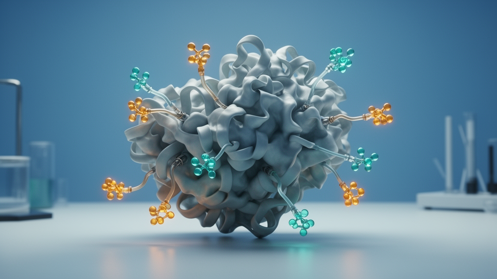
Perhaps the most fascinating layer of regulation comes from post-translational modifications, chemical tags that alter CYLD’s behavior without changing its genetic sequence. Phosphorylation, the addition of phosphate groups to serine or threonine residues, is the best studied. CYLD carries a cluster of phosphorylation sites between its second and third CAP-Gly domains, with serine 418 being a particularly important target. This residue is phosphorylated in response to a wide range of stimuli, including TNF, bacterial products, antigen receptor engagement, and constitutively in some aggressive blood cancers. The kinases responsible include the canonical IKKβ and the noncanonical TBK1 and IKKε. Intriguingly, the consequences of phosphorylation are disputed: some studies report that it dampens CYLD’s activity, promoting cell survival in T-cell leukemia, while others find that it enhances the enzyme’s ability to trim lysine 63 chains. A recent mass spectrometry analysis identified six phosphorylation sites in TNF-stimulated cells, five of which fall within the phospho-patch, suggesting that multiple modifications may act in concert rather than in isolation.
CYLD is also itself a target of ubiquitination, a poetic irony for an enzyme that spends its life removing ubiquitin from others. The E3 ligase SCFβ-TRCP ubiquitinates CYLD and reduces its abundance, a process that requires prior phosphorylation and plays a role in osteoclast differentiation. Another E3 ligase, MIB2, decorates CYLD with lysine 48 chains at two specific lysines, driving its proteasomal degradation and thereby amplifying NF-κB signaling in response to TNF. In hypoxic tumors, RNF213 ubiquitinates both CYLD and SPATA2, targeting them for lysosomal degradation and initiating pyroptotic cell death. Beyond ubiquitin, CYLD is modified by SUMOylation, the attachment of small ubiquitin-like modifiers. In neuroblastoma cells and glioblastoma stem-like cells, SUMOylation at lysine 40 impairs CYLD’s catalytic activity, leading to hyperactivation of NF-κB and, in the case of glioblastoma, promoting an aggressive phenotypic transition of the tumor cells.
Finally, CYLD can be irreversibly cut by proteases, a modification that permanently alters its function. Caspase-8 cleaves CYLD at aspartic acid 215 following TNF stimulation, inactivating the enzyme and blocking necroptosis in fibroblasts and macrophages. During influenza A virus infection, caspase-8-mediated cleavage of CYLD enhances innate antiviral immunity by preventing the deubiquitination of signaling proteins. The paracaspase MALT1, a caspase-like enzyme central to lymphocyte activation, cuts CYLD at arginine 324 in T cells, in aggressive B-cell lymphomas, and in glioblastoma stem cells, generally silencing the enzyme and promoting cell survival. Remarkably, recent work suggests that the N-terminal fragment released by caspase-8 cleavage is secreted into the bloodstream and may serve as a biomarker of septic shock. As the Nantes authors emphasize, mapping the complete landscape of CYLD’s modifications, understanding how they interact hierarchically, and exploring their interplay with SPATA2 represent the next great challenges for researchers hoping to exploit this remarkable enzyme therapeutically.
Subject of Research: Post-translational modifications regulating the deubiquitinating enzyme CYLD in cell death and inflammation
Article Title: The good, the bad, and the modified: CYLD’s post-translational tale
Article References: Macé, Y. M., Bidère, N., & Douanne, T. (2025). The good, the bad, and the modified: CYLD’s post-translational tale. Frontiers in Cell Death, 4, Article 1583221. https://doi.org/10.3389/fceld.2025.1583221
Image Credits: AI Generated
DOI: 10.3389/fceld.2025.1583221
Keywords: CYLD, deubiquitinase, post-translational modifications, NF-κB signaling, cell death, apoptosis, necroptosis, pyroptosis, SPATA2, phosphorylation, ubiquitination, tumor suppressor
Cite Scienmag News
Nathaniel Bowman. (October 16, 2026). The Tumor Suppressor CYLD: A Molecular Switchboard Controlled by Chemical Tags. Scienmag. https://scienmag.com/the-tumor-suppressor-cyld-a-molecular-switchboard-controlled-by-chemical-tags/
Nathaniel Bowman. "The Tumor Suppressor CYLD: A Molecular Switchboard Controlled by Chemical Tags." Scienmag, 16 October 2026, https://scienmag.com/the-tumor-suppressor-cyld-a-molecular-switchboard-controlled-by-chemical-tags/. Accessed 16 October 2026.
Nathaniel Bowman. "The Tumor Suppressor CYLD: A Molecular Switchboard Controlled by Chemical Tags." Scienmag. October 16, 2026. https://scienmag.com/the-tumor-suppressor-cyld-a-molecular-switchboard-controlled-by-chemical-tags/


