


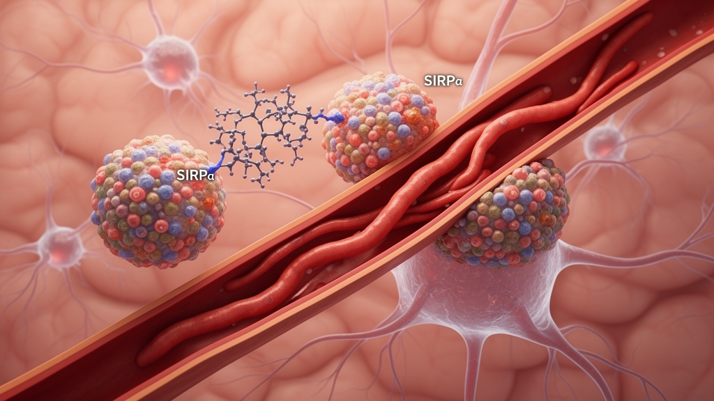
A preclinical study led by researchers at Wake Forest University School of Medicine identified SIRPα as a protein that may help triple-negative breast cancer cells spread to the brain and evade the local immune response. The investigators found that, inside these cancer cells, elevated SIRPα was associated with mitochondrial fission, greater cellular mobility, increased production of fibronectin and a weakened inflammatory response from brain-resident microglia. In laboratory experiments and preclinical models designed to reproduce breast-to-brain metastasis, reducing or inhibiting SIRPα slowed tumor growth, reduced the amount of cancer in the brain and delayed the development of brain metastases. The peer-reviewed findings were published in Neuro-Oncology on August 31, 2026, under the title “Cancer-intrinsic SIRPα Signaling Triggers Mitochondrial Fission and Immune Tolerance to Promote TNBC Breast-to-Brain Metastasis.” Because the work used human tumor data, cancer cells and preclinical models rather than a clinical trial, it does not establish that blocking SIRPα would improve outcomes in patients.
Triple-negative breast cancer, abbreviated TNBC, is an aggressive form of breast cancer that lacks three common markers used to guide treatment. According to the research summary, TNBC has a greater tendency than many other breast cancers to spread to the brain, where treatment options remain limited and outcomes are often poor. The study examined SIRPα, a protein best known for regulating immune cells, and found evidence that it can also act within TNBC cells. The investigators combined analysis of human breast cancer data and patient tumor samples with laboratory experiments involving cancer cells, immune cells and mitochondria, as well as preclinical models of breast cancer spread to the brain. This combination allowed them to connect a signal operating inside tumor cells with both changes in cancer-cell behavior and alterations in the surrounding brain environment.
The analysis of human material provided an initial association between SIRPα and TNBC. Researchers found higher levels of the protein in TNBC cells, particularly in tumors that had spread to the brain. Higher SIRPα levels were also associated with poorer outcomes among patients with TNBC. These observations did not by themselves demonstrate that SIRPα caused brain metastasis or poor outcomes in patients. Instead, the researchers used the patient findings to motivate mechanistic experiments in which they manipulated the protein in controlled systems. The broader study design therefore included clinical tumor material, cell-based experiments and animal models, with the models used to assess tumor growth, spread to the brain and changes in immune evasion under experimentally imposed conditions.
One central mechanism involved mitochondria, the structures that produce energy within cells. Laboratory experiments suggested that SIRPα changes mitochondrial behavior: in cancer cells with high levels of the protein, mitochondria broke into smaller pieces through a process called mitochondrial fission. This structural change was linked to increased cellular mobility and a greater likelihood of spreading in the study models. The experiments support a mechanistic relationship among cancer-cell SIRPα signaling, mitochondrial fission and metastatic behavior under the conditions tested. They do not establish that mitochondrial fission alone is sufficient to cause brain metastasis or that it is the only cellular process affected by SIRPα. Rather, the results place fission within a broader mechanism in which signaling inside a cancer cell alters energy-related structures and is associated with increased movement and metastatic capacity.
SIRPα also increased production of fibronectin, a protein that helps provide structure and support around cells. This finding extended the proposed mechanism beyond the internal behavior of the cancer cell to the microenvironment surrounding it. In the experimental systems, fibronectin appeared to weaken the response of microglia, the immune cells that help protect the brain. With repeated exposure to fibronectin, microglia became less able to trigger inflammation and attack cancer cells. The measurements therefore linked elevated SIRPα in tumor cells with both greater aggressive behavior and a local immune environment that appeared less able to respond. The investigators described this combination as helping cancer cells spread and “hide,” but the experiments do not show that microglia transport metastatic cells or that reduced inflammation is the only means by which TNBC cells colonize the brain.
When the investigators reduced or inhibited SIRPα across several preclinical systems, the consequences included slowed tumor growth, a decrease in the amount of cancer found in the brain and delayed development of brain metastases. Blocking the protein also reversed some of the changes associated with the cancer cells’ ability to evade the brain’s immune response. These intervention results provide experimental evidence that SIRPα contributes to the tumor-growth, metastatic and immune-related outcomes measured in the models, rather than appearing only as a marker of already aggressive tumors. At the same time, these systems do not reproduce every feature of human metastatic disease. The experiments investigate mechanism and target dependence; they do not demonstrate clinical efficacy, safety, an appropriate treatment schedule or a benefit for any particular group of patients.
Corresponding author David R. Soto-Pantoja, Ph.D., an associate professor of cancer biology at Wake Forest University School of Medicine, said the findings suggest that SIRPα helps make tumor cells more aggressive while changing the brain environment in ways that help those cells survive. He said the weakening of the brain’s immune-cell response was among the study’s most intriguing findings because it creates a more favorable environment for cancer cells to grow and survive. His observations are consistent with the reported experimental pattern: SIRPα-linked changes in mitochondria and fibronectin accompanied increased cell mobility, reduced microglial inflammatory responses and greater metastatic behavior. The study brings together several processes involved in brain metastasis, including cancer-cell energy production, the structural environment around cells and the response of brain-resident immune cells.
The results distinguish SIRPα from a protein that acts only as an immune-cell switch. Most experimental treatments involving the CD47-SIRPα pathway have focused on its role in immune cells, whereas these findings show that SIRPα can also act inside cancer cells. Targeting it could potentially interfere directly with processes within tumor cells that help them spread, in addition to affecting features of the immune environment. That therapeutic possibility remains a hypothesis for further testing rather than a demonstrated treatment. The investigators plan to continue studying how SIRPα functions in cancer cells, whether it can be safely targeted and whether blocking SIRPα could enhance existing immunotherapies or other treatments for TNBC brain metastases.
For patients with TNBC that has spread to the brain, or is at high risk of spreading, the study identifies a mechanism and a potential intervention that require further investigation. It does not provide evidence for changing current clinical care, selecting a drug or combining treatments. Brain metastases remain difficult to treat, and SIRPα was reduced or inhibited in controlled research settings rather than tested as a clinical therapy in people. Further studies must establish whether suppressing SIRPα produces consistent effects across tumors, what consequences blocking its normal biological functions may have, whether the approach can be delivered appropriately and whether the model results translate into clinical benefit. The study’s bounded conclusion is that, in preclinical systems, SIRPα signaling inside TNBC cells was linked to mitochondrial fission, increased motility, greater fibronectin production, weakened microglial responses and brain metastasis, while suppressing the protein produced antitumor and immune-related effects that merit further study.
Subject of Research: Preclinical investigation of SIRPα signaling in triple-negative breast cancer brain metastasis
Article Title: Researchers uncover how aggressive breast cancer may spread to the brain
Article References: Researchers uncover how aggressive breast cancer may spread to the brain. Original publication (Bibliographic details unavailable.)
Image Credits: AI Generated
DOI: Not provided
Keywords: SIRPα, triple-negative breast cancer, brain metastasis, cancer immunology, mitochondrial fission, fibronectin, microglia, tumor microenvironment, preclinical models, therapeutic target, Researchers, uncover
Cite Scienmag News
Nathaniel Bowman. (October 26, 2026). SIRPα promotes triple-negative breast cancer brain metastasis in models. Scienmag. https://scienmag.com/sirp%ce%b1-promotes-triple-negative-breast-cancer-brain-metastasis-in-models/
Nathaniel Bowman. "SIRPα promotes triple-negative breast cancer brain metastasis in models." Scienmag, 26 October 2026, https://scienmag.com/sirp%ce%b1-promotes-triple-negative-breast-cancer-brain-metastasis-in-models/. Accessed 26 October 2026.
Nathaniel Bowman. "SIRPα promotes triple-negative breast cancer brain metastasis in models." Scienmag. October 26, 2026. https://scienmag.com/sirp%ce%b1-promotes-triple-negative-breast-cancer-brain-metastasis-in-models/


