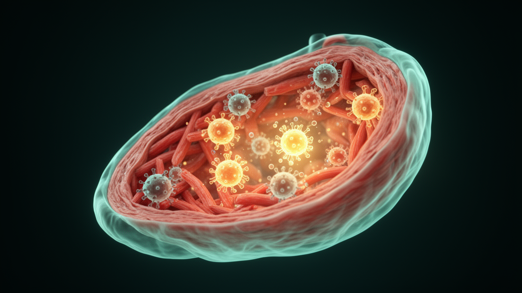
Viral myocarditis remains one of the most dangerous inflammatory heart diseases worldwide, disproportionately claiming the lives of children and young adults through sudden cardiac death, progressive heart failure and chronic dilated cardiomyopathy. A comprehensive review published in Experimental & Molecular Medicine now maps the cellular and molecular architecture of cardiac antiviral innate immunity from initial viral sensing through to therapeutic opportunity, synthesizing how these responses establish what the authors term the myocardial ‘inflammatory set point’ across both cardiac-resident cells and recruited innate immune populations. The work arrives at a moment when effective, mechanism-guided therapies for viral myocarditis remain conspicuously limited despite decades of experimental progress.
The true incidence of myocarditis is notoriously difficult to determine because the disease is often asymptomatic, with only approximately 10 percent of patients who have clinical evidence estimated to develop symptoms. Autopsy reports worldwide place incidence rates between 0.12 and 12 percent, while population-based estimates range from 4 to 14 per 100,000 people. Global burden data from the Global Burden of Disease study suggest that myocarditis accounted for roughly 1.27 million incident cases worldwide in 2019, with absolute numbers climbing over recent decades. Viral infections remain the leading cause, and the dominant etiologic agents vary across age, geographic region, season and diagnostic modality. Historically, enteroviruses—particularly coxsackievirus B3—have served as prototypical triggers in both experimental and clinical studies, but contemporary series increasingly emphasize that the leading viruses reported may shift over time, in part because viral detection depends on sampling method, assay sensitivity and the timing of specimen collection relative to infection and immune clearance.
Viral myocarditis is often conceptualized as a triphasic disease. The initial phase is driven by viral entry and replication in the heart, most notably in cardiomyocytes. This is followed by an immune activation phase in which innate and adaptive responses cooperate to limit viral spread but can simultaneously promote myocardial damage through excessive inflammation, cytotoxicity and autoimmune-like mechanisms. In the third phase, persistent injury and maladaptive repair contribute to fibrosis and ventricular remodeling, culminating in dilated cardiomyopathy. The review classifies cardiotropic viruses broadly by genome type. Typical RNA viruses include coxsackievirus B3, influenza viruses, human immunodeficiency virus, hepatitis C virus, encephalomyocarditis virus, reovirus, ECHO viruses and respiratory syncytial virus. Typical DNA viruses encompass cytomegalovirus, adenovirus, parvovirus B19, human herpesvirus 6 and Epstein–Barr virus. Recent evidence also supports SARS-CoV-2 as an infectious trigger in some patients during the COVID-19 pandemic, and viral myocarditis has recently been reported in association with Oz virus, a thogotovirus within the Orthomyxoviridae family.
Cardiac-resident cells are not passive targets of infection. Cardiomyocytes, the contractile muscle cells that constitute the major cellular mass of the myocardium, express multiple pattern-recognition receptors and stress-response pathways, produce cytokines and chemokines that recruit leukocytes, and can undergo regulated cell-death programs that shape both viral clearance and tissue injury. One cardiomyocyte-intrinsic pathway linked to viral myocarditis involves the tripartite motif family protein TRIM29, which engages the endoplasmic reticulum stress sensor PERK. Upon activation by ER stress, PERK autophosphorylates and phosphorylates eukaryotic translation initiation factor 2α, attenuating global translation while selectively promoting stress-responsive transcriptional programs. Sustained PERK–eIF2α signaling induces C/EBP homologous protein, a key mediator of pro-apoptotic signaling during prolonged ER stress. In a CVB3-induced mouse model, TRIM29 promotes PERK recruitment and enhances PERK activation—at least in part by facilitating PERK SUMOylation—thereby amplifying PERK–eIF2α–CHOP signaling, increasing cardiomyocyte apoptosis and worsening myocardial injury and disease progression. These observations support a broader concept: cardiomyocyte stress pathways are not merely downstream consequences of inflammation but can actively shape disease outcomes.
Cardiac fibroblasts, the principal stromal cells of the myocardium and the major source of extracellular matrix proteins, also serve as sentinel cells that rapidly respond to tissue stress by sensing pathogen-associated and damage-associated molecular patterns. In viral myocarditis, fibroblasts promote disease through three linked programs. They act as local inflammatory amplifiers, secreting TNF-α, IL-6 and MCP-1/CCL2 that recruit and instruct monocytes, macrophages and neutrophils. They shape the myocardial microenvironment by modulating endothelial activation, leukocyte retention and local cytokine gradients, creating feed-forward stromal–immune loops that aid early antiviral coordination but become maladaptive when prolonged. They drive remodeling and fibrosis by transitioning to myofibroblasts that deposit extracellular matrix, alter matrix composition and stiffness, and promote scar formation. Experimental work comparing CVB3 infection across murine cardiac cell types reported that fibroblasts not only mount robust inflammatory responses but may also show susceptibility to viral infection and replication, raising the possibility that infected or activated fibroblasts serve as local sources of inflammatory mediators that amplify leukocyte recruitment and exacerbate myocardial injury.
Among classical innate immune cells, natural killer cells occupy a particularly prominent position as front-line effectors across diverse viral etiologies. Unlike T cells, NK cells do not require prior sensitization to a specific viral peptide–MHC complex. Instead, they integrate signals from activating and inhibitory receptors to detect ‘altered-self’ states in infected or stressed cells, such as reduced MHC class I expression and increased expression of stress-induced ligands. In viral myocarditis, NK cells kill virus-infected cardiomyocytes to limit viral dissemination, and deficiency or depletion of NK cells in mouse models is associated with higher cardiac viral titers and increased myocardial injury. However, NK cells can also promote immunopathology when activation is excessive or prolonged. Reduced perforin activity attenuates tissue injury in encephalomyocarditis virus-induced mouse models, underscoring that NK-mediated cytotoxicity—while antiviral—can be a major driver of myocardial necrosis and subsequent remodeling if not properly regulated. Mechanistically, NK cells exert antiviral activity through direct cytotoxic killing via granule exocytosis of perforin and granzymes, death receptor pathways including FasL/Fas and TRAIL/TRAIL receptors, and cytokine-mediated effects through IFN-γ and TNF-α that suppress viral replication and enhance antigen presentation. Host factors further modulate NK cell impact: FOXO3 has been identified as a negative regulator of NK cell antiviral activity, and sex hormones influence NK cell responses, with estrogen dampening IFN-γ production and reduced estrogen associated with worsened myocarditis in females.
Dendritic cells bridge innate sensing, tissue inflammation and adaptive immunity through specialized subsets with distinct sensing and instructional roles. The cDC1 cells specialize in cross-presentation of exogenous antigens on MHC class I and efficient priming of cytotoxic CD8+ T cells; in the heart, CD103+ cDC1-mediated surveillance protects against progression of subclinical viral myocarditis. The cDC2 cells preferentially present antigen through MHC class II to CD4+ T cells, shaping their polarization toward inflammatory Th1 or Th17 effector programs or, in regulatory contexts, toward regulatory T cells that restrain inflammation. Plasmacytoid dendritic cells serve as rapid and robust producers of type I interferon following endosomal sensing of viral nucleic acids. Beyond canonical antigen presentation, the review highlights PARP9 as a noncanonical, MAVS-independent RNA sensor in dendritic cells that binds viral double-stranded RNA and promotes type I interferon responses through a PI3K–AKT3 signaling axis leading to phosphorylation and activation of IRF3 and IRF7. PARP9-deficient mice exhibit increased susceptibility to reovirus-induced viral myocarditis due to impaired type I interferon responses, illustrating that RNA sensing in this disease is broader than the classical TLR/RLR framework.
Macrophages constitute a major fraction of infiltrating leukocytes in inflamed myocardium and participate in viral sensing, phagocytosis, cytokine production, antigen presentation, efferocytosis and tissue repair. Contemporary single-cell and spatial transcriptomic studies show that cardiac macrophages occupy a spectrum of states shaped by ontogeny, tissue niche, viral load, cytokines and time after injury, including resident cardiac CCR2− macrophages, recruited CCR2+ monocyte-derived inflammatory macrophages, IFN-stimulated macrophages, antigen-presenting macrophages and reparative or profibrotic macrophage states. Macrophage-intrinsic signaling through type I interferon production pathways, inflammasomes and cytokine receptors tunes the balance between type I interferon-mediated viral restriction and heart injury driven by proinflammatory cytokines IL-1β, TNF-α and IL-6. The E3 ubiquitin ligase TRIM18 recruits protein phosphatase 1A to dephosphorylate TANK-binding kinase 1, thereby dampening RNA sensing and MAVS-dependent type I interferon signaling and increasing the severity of CVB3-induced cardiac inflammation. Neutrophils, the most abundant circulating leukocytes in humans, are recruited within minutes to hours to sites of infection or sterile tissue injury, where they eliminate viruses through phagocytosis, degranulation of antimicrobial proteins, generation of reactive oxygen species and formation of neutrophil extracellular traps. PAD4-knockout mice unable to form these traps showed lower monocyte presence, reduced proinflammatory signals and decreased cardiac necrosis in both early and late acute viral myocarditis, suggesting that NET formation is detrimental to cardiac tissue during acute disease.
On the molecular sensing level, the review dissects RNA- and DNA-sensing pathways with technical precision. Endosomal TLR3 recognizes viral double-stranded RNA and signals through the adaptor TRIF, which promotes IRF activation and antiviral interferon programs; TRIF-deficient mice infected with CVB3 exhibit higher viral titers, worse cardiac dysfunction and increased early mortality. TLR7 and TLR8 detect single-stranded RNA within endosomes, and exposure to CVB3 ssRNA upregulates both receptors, driving inflammatory cytokine production in a TLR7/8-dependent manner. In the cytosol, RIG-I-like receptors detect viral RNA and signal through the mitochondrial adaptor MAVS, triggering downstream activation of IRF3/IRF7 and NF-κB. RIG-I responds strongly to shorter dsRNA and 5′-triphosphorylated ssRNA, whereas MDA5 is preferentially activated by longer dsRNA and is particularly important for picornaviruses such as CVB3. In encephalomyocarditis virus infection, MDA5 or MAVS deficiency results in markedly increased viral titers in the heart early after infection. For DNA viruses, the cytosolic sensor cGAS binds double-stranded DNA in a sequence-independent manner and produces cGAMP, which activates STING on the endoplasmic reticulum, leading to recruitment of TBK1, phosphorylation of IRF3 and induction of type I interferon alongside NF-κB-dependent proinflammatory cytokines. DNA viruses frequently antagonize cGAS–STING: Epstein–Barr virus can induce TRIM29 to target STING for degradation, reducing type I interferon output. The AIM2 inflammasome, upon binding cytosolic dsDNA, recruits the adaptor ASC to activate caspase-1, which processes proIL-1β and proIL-18 into mature IL-1β and IL-18 and cleaves gasdermin D to generate its pore-forming N-terminal fragment, promoting cytokine secretion and inflammasome-associated pyroptotic cell death.
Translational efforts increasingly recognize that therapeutic benefit depends on etiology and disease phase. Core clinical biomarkers include cardiac troponins reflecting cardiomyocyte injury, BNP/NT-proBNP indicating myocardial wall stress, and C-reactive protein supporting an inflammatory phenotype. Cardiac magnetic resonance imaging provides noninvasive tissue-level biomarkers including edema and injury or fibrosis measures. Endomyocardial biopsy with histology, immunostaining and viral genome testing remains the most direct approach to classify myocarditis as virus-positive versus virus-negative inflammatory cardiomyopathy, a stratification that is clinically consequential because immunosuppression can be detrimental during active viral replication. Liquid biopsy strategies using circulating viral genomes, cell-free DNA and microRNAs may complement biopsy and cardiac MRI for real-time risk stratification. In COVID-19-associated viral myocarditis, transcriptomic analysis demonstrated enrichment of JAK–STAT3, inflammatory and interferon-response gene sets, with B2M and C3 proposed as diagnostic markers. Preclinical strategies include type I interferon augmentation for viral persistence, NLRP3 inflammasome inhibition with MCC950, platelet-derived extracellular vesicle immunomodulation, and pharmacologic PERK inhibition with GSK2656157, which mitigated disease in CVB3-induced mouse models by disrupting the TRIM29–PERK connection. Clinical trials now evaluate interferon-β-1b for chronic viral cardiomyopathy, anakinra for IL-1 pathway blockade in acute myocarditis, and colchicine across the myocarditis and myopericarditis spectrum. The review proposes a phase- and etiology-guided framework pairing antiviral immunomodulation with timely restraint of innate proinflammatory amplifiers to limit heart damage and prevent progression from acute viral myocarditis to dilated cardiomyopathy and heart failure, while also highlighting the emerging gut–heart immune axis, in which intestinal barrier integrity, commensal microbiota and mucosal innate immune tone can influence viral burden, systemic cytokine responses and subsequent cardiac inflammation.
Subject of Research: Antiviral innate immune mechanisms and emerging therapeutics in viral myocarditis
Article Title: Antiviral innate immunity in myocarditis: mechanisms and emerging therapeutics
Article References: Hammons, C., Mohanram, A., Nguyen, P., Lu, W., Jacobs, B., Li, X. C., Zhang, Z., & Xing, J. (2026). Antiviral innate immunity in myocarditis: mechanisms and emerging therapeutics. Experimental & Molecular Medicine, 58(9), 2825-2843. https://doi.org/10.1038/s12276-026-01822-z
Image Credits: AI Generated
DOI: 10.1038/s12276-026-01822-z
Keywords: viral myocarditis, innate immunity, pattern-recognition receptors, type I interferon, inflammasome, cardiomyocytes, natural killer cells, dendritic cells, macrophages, cGAS-STING, TLR signaling, heart failure
Cite Scienmag News
Kristina Jarvis. (October 24, 2026). How Antiviral Innate Immunity Shapes Viral Myocarditis Outcomes. Scienmag. https://scienmag.com/how-antiviral-innate-immunity-shapes-viral-myocarditis-outcomes/
Kristina Jarvis. "How Antiviral Innate Immunity Shapes Viral Myocarditis Outcomes." Scienmag, 24 October 2026, https://scienmag.com/how-antiviral-innate-immunity-shapes-viral-myocarditis-outcomes/. Accessed 24 October 2026.
Kristina Jarvis. "How Antiviral Innate Immunity Shapes Viral Myocarditis Outcomes." Scienmag. October 24, 2026. https://scienmag.com/how-antiviral-innate-immunity-shapes-viral-myocarditis-outcomes/