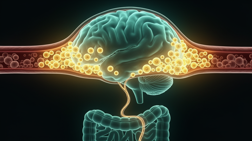
Infections in the gut can leave a lasting imprint on the immune defenses of the brain, according to a study in mice published in Nature Neuroscience. Researchers led by Menna Clatworthy at the University of Cambridge found that orally administered bacterial and parasitic infections reshape the population of CD4+ T cells in the dura mater, the tough outermost layer of the meninges, seeding long-lived, antigen-specific memory T cells that persist for months and can mount rapid recall responses when the same pathogen later appears in the bloodstream.
The dura mater forms the border between the brain and the rest of the body and houses a rich network of immune cells. Its vasculature is fenestrated, meaning it contains small openings that allow bloodborne microorganisms to reach the central nervous system. In humans, the organisms most commonly found in the blood of people with bacteremia are gut-derived Gram-negative bacteria, yet Gram-negative meningitis remains infrequent, suggesting that effective defense mechanisms exist at this border. The new work identifies one such mechanism: a direct cellular link between intestinal and dural immunity.
In steady-state conditions, the team found that CD4+ T cells formed a minority of immune cells in the dura and were predominantly non-naive, with around half expressing the transcription factor T-bet. To test whether intestinal challenges could alter this population, the researchers used several mouse models. When mice received dextran sulfate sodium, or DSS, a chemical that breaches the intestinal epithelial barrier and drives expansion of T helper 17 cells in the colon, the team observed a matching increase in TH17 cells in the extravascular compartment of the dura, in aggregates around the dural venous sinuses and in a dural lymphoid structure called rostral-rhinal DALT, with no significant change in the spleen or in the intravascular dural compartment.
A similar pattern followed oral infection with Citrobacter rodentium, a bacterium known to induce intestinal TH17 responses. Twenty-one days after infection, both colonic and dural RORγt-expressing CD4+ T cells had expanded significantly, and the dural TH17 cells were predominantly producers of the cytokine IL-17. Spatial imaging confirmed that these cells sat along the dural venous sinus and within the rostral-rhinal DALT. Notably, an attenuated mutant strain of C. rodentium that induces less intestinal IL-22 secretion and neutrophil recruitment produced a similar level of dural TH17 expansion, supporting the idea that the cells were activated in the intestine and then relocated.
The polarization of dural T cells tracked the type of intestinal stimulus. When mice were infected with the parasitic worm Schistosoma mansoni, which drives a strong TH2 response in the gut, the researchers found a marked expansion of GATA-3-expressing TH2 cells in both intestine and dura 42 days later, with confocal imaging showing increased CD4+ T cells in aggregates along the venous sinus walls. By contrast, oral infection with Trichuris muris, the mouse whipworm, which induces a dominant TH1 response in C57BL/6 mice, led to an increase in T-bet-expressing TH1 cells in both colon and dura. Together, these results show that the nature of an intestinal infection determines not only gut T cell polarization but also the polarization of tissue-resident dural CD4+ T cells.
To probe whether the gut and dural T cells were related, the team performed T cell receptor sequencing on paired samples of dura and intestinal segments after DSS colitis. They found a significant increase in identical T cell receptors shared between dura and duodenum and distal colon in colitis mice compared with controls, suggesting that clones expanded in the intestine seed the dura. Using an attenuated Salmonella enterica serovar Typhimurium engineered to carry a known antigenic peptide called 2W1S, they confirmed that antigen-specific CD4+ T cells expanded in the extravascular compartments of both colon and dura 21 days after oral infection. These dural cells expressed the tissue-residency marker CD69, showed an effector memory phenotype and secreted IFN-γ. Gut-derived antigen-specific cells were also found in the liver, lung, kidney and bladder, hinting that gut-to-organ immune seeding may be a broader strategy.
The migration route depended on a specific chemokine axis. Single-cell RNA sequencing data showed that Cxcr6 was the most highly expressed chemokine receptor on gut CD4+ T cells regardless of the polarizing stimulus, while its ligand Cxcl16 was among the most highly expressed chemokine transcripts in the dura, produced mainly by meningeal macrophages. When the researchers blocked CXCL16 with a neutralizing antibody after oral Salmonella challenge, the number of antigen-specific CD4+ T cells recruited to the dura fell, without affecting the colon or spleen. Conversely, in mixed bone marrow chimeras, CXCR6-deficient T cells were recruited to the dura far less efficiently than normal cells, confirming that the CXCR6-CXCL16 axis mediates the relocation.
Crucially, the relocated cells behaved as genuine memory cells. Five and a half weeks after oral priming with the 2W1S-expressing Salmonella, mice that received an intravenous rechallenge showed a robust expansion of dural antigen-specific CD4+ T cells, whereas cells were almost undetectable in mice given the intravenous challenge alone. Remarkably, the recall response still worked five months after the initial oral infection. Single-cell analysis showed these cells expressed memory and tissue-residency markers, including CD69 and the integrin VLA-4, which is known to be important for lymphocyte entry into the central nervous system. Experiments with dural wholemounts stimulated with the 2W1S peptide confirmed that the resident dural cells themselves, rather than newly arrived circulating cells, recognized the antigen and activated, showing Nur-77 signaling and IFN-γ production.
The memory cells also appeared to contribute to brain defense. Mice orally primed with the 2W1S-expressing Salmonella and later challenged intravenously with a virulent strain showed significantly fewer bacteria in the brain than unprimed controls, and the number of dural antigen-specific cells correlated negatively with brain bacterial counts. Because the researchers could not selectively deplete dural CD4+ T cells without depleting them systemically, they used a stringent cross-protection model in which the only shared antigen between oral priming and intravenous challenge was the single 2W1S peptide: mice primed orally with 2W1S-expressing C. rodentium and later challenged with 2W1S-expressing Candida albicans showed reduced fungal loads in the brain, an expansion of antigen-specific CD4+ T cells in both dura and brain parenchyma, and reduced microglial activation, although the protective effect was modest.
The findings carry implications beyond infection. Because meningeal T cells can influence brain function through cytokine production, and because gut-derived IL-17-secreting TH17 cells expanded in the dura during a model of inflammatory bowel disease, the authors suggest that such cells might contribute to the elevated rates of anxiety and depression seen in patients with IBD, conditions reported in up to 30 percent and 25 percent of these patients respectively. The work also raises questions about chronic schistosomiasis, which affects around 140 million people and was associated here with substantial dural TH2 expansion. More broadly, the study proposes that migratory immune cells act as an information relay between peripheral organs and the brain, using cytokines rather than neurotransmitters, allowing the central nervous system to accumulate immunological memory of the microorganisms most likely to reach it through the blood.
Subject of Research: How intestinal infections establish long-lived memory CD4+ T cell populations in the brain's dural meninges
Article Title: Intestinal infections establish antigen-specific, long-lived memory CD4+ T cells in the brain and meninges
Article References: Fleming, A., Neish, K., Di Marco-Barros, R., Posner, D. A., Lee, C. Y. C., Stewart, A., Tuong, Z. K., Bremridge, M., Peñalver, A., Cabantous, M., Richoz, N., Portet, A., Harcourt, K., Gillman, E., Sow, T. T. M., Hasegawa, T., Ruano-Gallego, D., Frankel, G., Withers, D., … Clatworthy, M. R. (2026). Intestinal infections establish antigen-specific, long-lived memory CD4+ T cells in the brain and meninges. Nature Neuroscience. https://doi.org/10.1038/s41593-026-02428-4
Image Credits: AI Generated
DOI: 10.1038/s41593-026-02428-4
Keywords: meninges, dura mater, CD4+ T cells, intestinal infection, immunological memory, CXCR6-CXCL16, TH17 cells, Salmonella, schistosomiasis, gut-brain immunity, Nature Neuroscience, mouse study
Cite Scienmag News
Cassandra Pierce. (October 24, 2026). Gut Infections Leave Long-Lasting Immune Memory at the Brain’s Border. Scienmag. https://scienmag.com/gut-infections-leave-long-lasting-immune-memory-at-the-brains-border/
Cassandra Pierce. "Gut Infections Leave Long-Lasting Immune Memory at the Brain’s Border." Scienmag, 24 October 2026, https://scienmag.com/gut-infections-leave-long-lasting-immune-memory-at-the-brains-border/. Accessed 24 October 2026.
Cassandra Pierce. "Gut Infections Leave Long-Lasting Immune Memory at the Brain’s Border." Scienmag. October 24, 2026. https://scienmag.com/gut-infections-leave-long-lasting-immune-memory-at-the-brains-border/