Copper is one of biology’s most essential trace elements, serving as a cofactor for enzymes that drive mitochondrial respiration, antioxidant defense, and the synthesis of hormones, neurotransmitters, and pigments. Yet the same metal that sustains life can destroy it. A new review published in Frontiers in Cell Death by Yujuan Jiao, Hongxin Wang, and colleagues at Harbin Medical University synthesizes the rapidly expanding field of copper-dependent cell death, arguing that the metal’s dual nature offers one of the most promising and unexpected avenues in modern cancer research. The work arrives at a moment when scientists have only just begun to map the molecular machinery of a phenomenon that was formally named just three years ago.
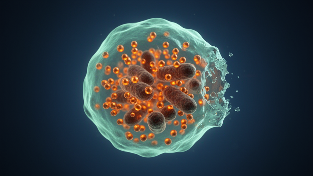
In 2022, Peter Tsvetkov and colleagues first proposed the concept of “cuproptosis,” a copper-dependent, regulated form of cell death that is fundamentally distinct from apoptosis, necroptosis, pyroptosis, and ferroptosis. The defining feature of cuproptosis is its reliance on mitochondrial respiration. When excess copper accumulates inside a cell, it binds directly to lipoylated components of the tricarboxylic acid (TCA) cycle, the metabolic engine that converts nutrients into usable energy. Lipoylation is a post-translational modification in which an acyl group is covalently attached to lysine residues, and it occurs in only four key metabolic enzymes: DBT, DLST, DLAT, and GCSH. All four funnel carbon sources into the TCA cycle, making them linchpins of mitochondrial metabolism.
The consequences of copper binding to these lipoylated proteins are catastrophic. The metal induces abnormal oligomerization and aggregation of the proteins, generating severe proteotoxic stress. Simultaneously, copper destabilizes iron-sulfur (Fe-S) cluster proteins, essential cofactors involved in mitochondrial energy production, amino acid synthesis, and tRNA modification. The result is irreversible cell death that proceeds even when cells are engineered to lack the caspase-3 enzyme, a hallmark of apoptosis, or when they are treated with inhibitors of other known death pathways. This resistance to conventional blockers is what convinced researchers that cuproptosis represents an entirely new category of regulated cell death.
At the center of this process sits a small mitochondrial protein called ferredoxin 1, or FDX1. Under normal conditions, FDX1 transfers electrons from NADPH to the mitochondrial cytochrome P450 system and participates in steroid synthesis, cholesterol metabolism, and bile acid production. In the context of copper overload, however, FDX1 performs a far more consequential role: it reduces copper from its Cu2+ form to the more reactive Cu+ form, which then binds to lipoylated TCA cycle proteins such as DLAT. FDX1 also functions upstream of lipoic acid synthetase (LIAS), promoting Fe-S cluster assembly and maintaining the stability needed for protein lipoylation. Genetic studies have shown that deleting FDX1 or LIAS inhibits copper-induced cell death entirely, while FDX1 knockout leaves cells resistant to the copper ionophore elesclomol but unaffected by apoptosis inducers or ferroptosis agents.
Copper’s toxicity extends beyond lipoylated proteins to the Fe-S cluster assembly pathway itself. Excessive copper competes with iron for metal-binding sites in assembly proteins such as ISCA1, ISCA2, and ISCU, hindering the biosynthesis of both [2Fe-2S] and [4Fe-4S] clusters. When Fe-S cluster production falters, Fe-S apoproteins fail to mature properly and are flagged as misfolded, then degraded by the ubiquitin-proteasome system. The drop in Fe-S cluster availability also reduces LIAS levels, further suppressing protein lipoylation and compounding the metabolic collapse. Remarkably, non-Fe-S proteins remain largely unaffected, indicating that this mechanism is highly specific to copper’s interference with iron-sulfur chemistry.
The regulation of intracellular copper is itself a tightly choreographed affair. Dietary copper enters through the small intestine via the transporter CTR1, is exported across the basolateral membrane by ATP7A, and is excreted into bile through ATP7B, the protein whose mutations cause Wilson’s disease. Inside cells, a network of metallochaperones including COX17, CCS, and ATOX1 delivers copper to precise destinations, while metallothioneins and the small molecule glutathione buffer free copper levels. Under normal conditions, the intracellular concentration of free Cu2+ is estimated to be as low as 10 to the minus 18 molar, an extraordinary feat of homeostatic control. When the import gene SLC31A1 is overexpressed, cells become dramatically more sensitive to copper, showing reduced mitochondrial respiratory proteins, diminished lipoylation, declining Fe-S cluster proteins, and elevated heat shock protein 70, all signatures of impending cuproptosis.
What makes cuproptosis especially compelling for oncology is that cancer cells are uniquely vulnerable to it. Tumors exhibit significantly elevated copper levels in both tissue and serum across thyroid, breast, lung, pancreatic, gallbladder, and colorectal cancers, and this elevation correlates with poor prognosis. Cancer cells upregulate CTR1 to meet their heightened copper demand, redirect chaperone proteins toward proliferation- and angiogenesis-promoting enzymes, and activate signaling cascades such as MEK/ERK and BRAF that drive growth, VEGF-mediated angiogenesis, and epithelial-mesenchymal transition. Hypoxia-inducible factor-1α further stabilizes copper metabolism genes, creating a positive feedback loop. Yet this copper dependence is a double-edged sword: the same metal that fuels malignancy can be weaponized to destroy it, particularly because cancer cells already operate at higher basal oxidative stress and rely more heavily on mitochondrial metabolism than their healthy counterparts.
The copper ionophore elesclomol has emerged as the leading experimental tool for exploiting this vulnerability. Elesclomol binds extracellular Cu2+ through its nitrogen and sulfur atoms, forms a neutral complex, and delivers it directly into mitochondria, where FDX1 reduces the copper to its reactive Cu+ form. The ionophore is then exported and recycled, continuously shuttling more copper into the cell until antioxidant defenses are overwhelmed. Because normal cells possess robust copper homeostasis, intact mitochondria, and potent antioxidant systems, elesclomol-copper complexes are rarely activated in healthy tissue, minimizing collateral damage. The drug has shown activity against lung cancer, melanoma, and sarcoma, and has even been repurposed to treat Menkes disease, a neurological disorder of copper deficiency. Notably, copper delivery also upregulates PD-L1 on tumor cell surfaces, creating a rationale for combining cuproptosis induction with anti-PD-L1 immunotherapy.
Recent nanotechnology efforts have pushed this concept toward clinical translation. Researchers designed an amphiphilic biodegradable polymer nanoparticle, NP@ESCu, that encapsulates both elesclomol and copper and releases them in response to reactive oxygen species. The nanoparticle dramatically enhances intracellular copper transport, causes severe mitochondrial damage including shrinking, increased membrane density, and loss of cristae, and ultimately triggers cuproptosis. Beyond direct tumor killing, NP@ESCu promotes dendritic cell maturation, encourages CD8-positive T cell infiltration, and reprograms tumor-associated macrophages from the immunosuppressive M2 phenotype toward the pro-inflammatory M1 state, activating systemic anti-tumor immune responses and long-term immune memory. When combined with αPD-L1 antibodies, the strategy significantly improves the tumor immune microenvironment and therapeutic efficacy, offering a blueprint for next-generation copper-targeted cancer drugs.
Significant questions remain before cuproptosis can be fully harnessed in the clinic. Researchers still do not know whether a specific copper concentration threshold separates mere toxicity from true cuproptosis, or how the two states interact. Tumors have evolved resistance mechanisms, including glutathione-mediated copper chelation and HIF-1α-driven upregulation of metallothionein MT2A and degradation of DLAT under hypoxic conditions, which may be reversible with HIF-1α inhibitors. Biomarkers such as plasma ceruloplasmin activity and the serum copper-to-zinc ratio may help identify patients most likely to benefit. The tumor suppressor p53 adds another layer of complexity, potentially sensitizing cells to cuproptosis by suppressing glycolysis, promoting oxidative phosphorylation through SCO2 and AIF, and modulating glutathione biosynthesis. As the Harbin Medical University team concludes, systematic research into cuproptosis mechanisms, drug development, and clinical translation could open entirely new avenues for treating cancers that have long evaded conventional therapies.
Subject of Research: Copper-induced cell death mechanisms and their role in the tumor microenvironment
Article Title: Copper-induced cell death mechanisms and their role in the tumor microenvironment
Article References: Jiao, Y., Wang, H., Zhu, M., Liu, T., Li, Y., Yang, S., Zhang, M., & Zhang, L. (2025). Copper-induced cell death mechanisms and their role in the tumor microenvironment. Frontiers in Cell Death, 4, Article 1628470. https://doi.org/10.3389/fceld.2025.1628470
Image Credits: AI Generated
DOI: 10.3389/fceld.2025.1628470
Keywords: cuproptosis, copper, cell death, tumor microenvironment, FDX1, protein lipoylation, mitochondria, elesclomol, cancer therapy, immunotherapy, iron-sulfur clusters, PD-L1
Cite Scienmag News
Nathaniel Bowman. (October 17, 2026). Copper Overload Triggers a Distinct Form of Cell Death That Could Transform Cancer Therapy. Scienmag. https://scienmag.com/copper-overload-triggers-a-distinct-form-of-cell-death-that-could-transform-cancer-therapy/
Nathaniel Bowman. "Copper Overload Triggers a Distinct Form of Cell Death That Could Transform Cancer Therapy." Scienmag, 17 October 2026, https://scienmag.com/copper-overload-triggers-a-distinct-form-of-cell-death-that-could-transform-cancer-therapy/. Accessed 17 October 2026.
Nathaniel Bowman. "Copper Overload Triggers a Distinct Form of Cell Death That Could Transform Cancer Therapy." Scienmag. October 17, 2026. https://scienmag.com/copper-overload-triggers-a-distinct-form-of-cell-death-that-could-transform-cancer-therapy/