


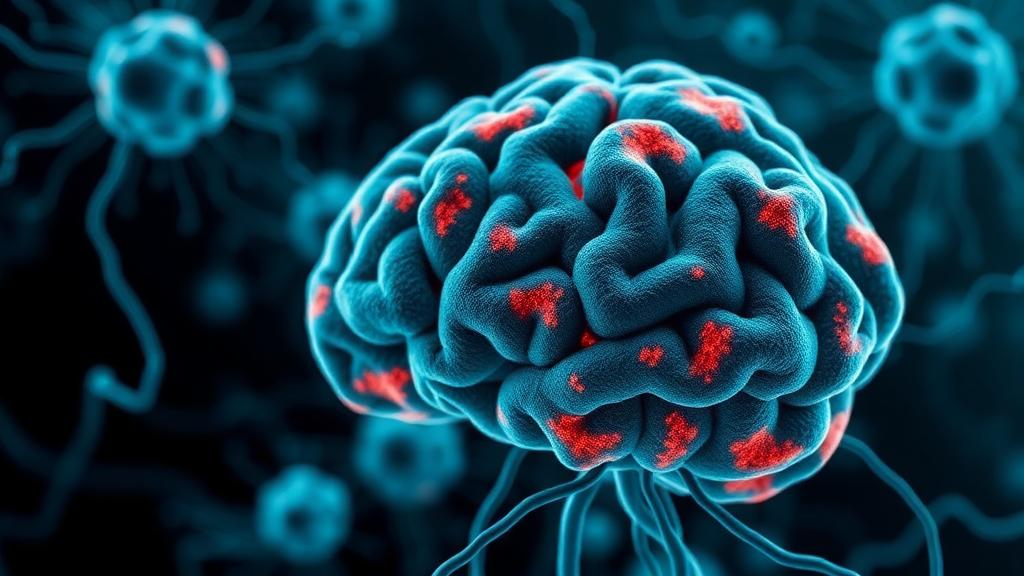
In a discovery that reframes how scientists understand the brain’s immune response in Parkinson’s disease, researchers at the University of Oxford have shown that microglia, the resident immune cells of the central nervous system, can actively strip toxic protein aggregates out of living neurons without destroying them. The finding, published in Science Translational Medicine and funded by the Medical Research Council and the National Institute for Health and Care Research Biomedical Research Centre in Oxford, reveals a protective mechanism that had gone entirely unnoticed in decades of research into the neurodegenerative disorder. Rather than standing by as dopamine-producing neurons accumulate the misfolded alpha-synuclein protein that defines Parkinson’s pathology, a specialised population of human microglia appears to perform a kind of cellular surgery, carefully excising the harmful material while leaving the neuron itself intact and functional.
The significance of this work lies in the central mystery it helps to address. Parkinson’s disease affects more than ten million people worldwide and is characterised by two intertwined processes: the progressive death of dopamine-producing neurons in a region of the brain called the substantia nigra, and the build-up of abnormal clumps of alpha-synuclein inside nerve cells. These aggregates, known as Lewy bodies and Lewy neurites at later stages, are thought to contribute to neuronal dysfunction and death. For years, therapeutic strategies have sought ways to prevent the formation of these clumps or to clear them once they appear, but the tools of the brain’s own immune system have generally been viewed with ambivalence. Microglia, when persistently activated, are known to release inflammatory signals that can damage neurons, and much of the recent literature has cast them as accomplices in neurodegeneration rather than as protectors.
The Oxford team, led by first author Dr Hung-Ju Chueh and senior author Professor George Tofaris of the Nuffield Department of Clinical Neurosciences, approached the question with a human-specific experimental system. They derived induced pluripotent stem cells, which can be reprogrammed to become almost any cell type in the body, and used them to generate both dopamine-producing neurons and microglia in the laboratory. This allowed the two cell types to be studied together in a controlled environment that reflects human biology far more closely than animal models, in which alpha-synuclein aggregation and microglial behaviour can differ substantially from the human condition. The researchers then created conditions in which alpha-synuclein aggregates formed inside the neurons, either by increasing the dosage of the alpha-synuclein gene or by exposing the cells to pre-formed alpha-synuclein fibrils that act as molecular templates, coaxing the neurons’ own protein to misfold in the same characteristic way.
What the team observed when microglia encountered these aggregate-laden neurons was striking. The immune cells did not engulf and digest entire neurons, as might be expected in a conventional phagocytic response to damaged cells. Instead, they engaged in a process called trogocytosis, from the Greek word for gnaw, in which one cell nibbles small portions of material from the membrane or cytoplasm of another. In this case, the microglia selectively removed the portions of the neuron containing the aggregated alpha-synuclein, effectively extracting the pathological protein from within the living cell. Dr Chueh emphasised the precision of this response, noting that the microglia were not simply engulfing damaged neurons but instead removing parts of the neuron containing aggregated alpha-synuclein, which suggests that at certain stages of the disease microglia help neurons dispose of potentially harmful material.
This distinction matters enormously for how the immune system is understood in neurodegeneration. Trogocytosis has been described in other contexts, particularly in the immune system’s interactions with tumour cells and in certain developmental processes, but its role in clearing intracellular protein aggregates from neurons had not been demonstrated before. The mechanism offers a way for the brain to deal with a problem that ordinary protein degradation pathways inside the neuron apparently cannot solve on their own. Aggregated alpha-synuclein is notoriously resistant to the cell’s internal disposal machinery, including the proteasome and lysosome systems, and once seeds of misfolded protein take hold they can propagate from cell to cell, spreading pathology through connected brain circuits. A mechanism by which microglia can reach into neurons and remove these seeds while preserving neuronal structure and function represents a fundamentally different kind of intervention from simply killing and replacing damaged cells.
The research went further, dissecting the molecular signals that govern this protective behaviour. The team found that the response was fine-tuned by communication between the two cell types through specific receptors: P2RY12, a receptor involved in microglial sensing and surveillance, and CD22, an inhibitory receptor that restrains immune activation. In addition, the microglia operated under what the researchers describe as a self-regulated brake, mediated by the cytokine IL-10, an anti-inflammatory signalling molecule that the cells produce themselves. This internal braking system prevents the microglia from escalating into a full inflammatory response that would damage healthy tissue, ensuring that the nibbling remains targeted and proportionate. The interplay of sensing signals, inhibitory checkpoints and self-produced anti-inflammatory feedback suggests a carefully calibrated system rather than a crude immune reaction.
Using single-cell RNA sequencing, a technique that allows researchers to read the gene expression profiles of individual cells rather than averaging across a whole population, the team identified a distinct subpopulation of activated microglia associated with this beneficial clearance response. This is an important methodological advance, because bulk analyses of microglial gene expression in diseased brains have often lumped together cells performing very different functions, some harmful and some protective. Being able to distinguish a clearance-competent microglial state opens the possibility of identifying equivalent cells in human brain tissue and, ultimately, of finding ways to expand or enhance this population therapeutically. Professor Tofaris noted that the findings highlight that the immune response in Parkinson’s disease is more nuanced than simply being beneficial or harmful, and that understanding how to enhance and monitor such beneficial microglial functions without triggering damaging inflammation could open up new avenues for developing disease-modifying treatments.
A second major discovery concerns a protein called glycoprotein non-metastatic melanoma protein B, or GPNMB, which appears to be an essential component of the protective machinery. Genetic variation at the GPNMB locus had previously been linked to Parkinson’s disease risk through genome-wide association studies, large-scale genetic surveys that compare the DNA of patients and healthy controls, but the functional role of the protein in the disease had remained unclear. The Oxford researchers found that GPNMB was upregulated in microglia exposed to neurons containing alpha-synuclein aggregates, and that the protein interacted with the pathological alpha-synuclein inside the microglia after the material had been nibbled away from the neurons. Crucially, when the team selectively reduced GPNMB expression in microglia using CRISPR interference, a genetic technique that silences specific genes without cutting the DNA, the cells became markedly less effective at clearing the aggregates, providing direct evidence that GPNMB plays an active role in the process rather than being a passive byproduct.
The human relevance of the GPNMB finding was reinforced by evidence from brain tissue. The researchers examined post-mortem tissue from the substantia nigra, the brain region most affected in Parkinson’s disease, from individuals who had died with either incidental alpha-synuclein pathology, an asymptomatic stage in which the protein aggregates are present without clinical disease, or full Parkinson’s disease. In both cases, GPNMB was increased in the microglia of this vulnerable region. This pattern suggests that the protective response is not merely an artefact of the laboratory system but occurs in the human brain, and that it may be engaged even in the earliest pathological stages, before symptoms emerge. If so, the window during which enhancing microglial clearance could slow or prevent disease progression may be wider than previously assumed, and GPNMB levels could potentially serve as a marker of this protective activity.
The broader implications of the study extend across several fronts in Parkinson’s research. First, it provides a concrete mechanism by which the immune system can be protective in neurodegeneration, countering the prevailing narrative of microglia as primarily inflammatory villains and suggesting that blanket immunosuppression might inadvertently remove a beneficial defence. Second, it identifies molecular targets, including the P2RY12 and CD22 signalling pathways, the IL-10 brake and GPNMB itself, that could be manipulated pharmacologically to strengthen the clearance response. Third, it demonstrates the power of human stem cell co-culture systems combined with single-cell profiling to reveal biology that animal models have missed. Much work remains before these findings translate into treatments: the researchers and the field will need to determine how trogocytosis is triggered in the living brain, how it changes as disease advances, and whether enhancing it in patients is safe and effective. But for a disease in which available therapies manage symptoms without slowing the underlying neurodegeneration, the image of brain immune cells carefully nibbling toxic protein out of struggling neurons offers a genuinely new and hopeful direction for disease-modifying drug development.
Subject of Research: Microglial clearance of intraneuronal alpha-synuclein aggregates in Parkinson's disease
Article Title: Brain immune cells protect neurons in Parkinson’s disease by “nibbling” harmful material
Article References: Brain immune cells protect neurons in Parkinson’s disease by “nibbling” harmful material. (n.d.). Original publication
Image Credits: AI Generated
DOI: Not provided
Keywords: Parkinson's disease, microglia, alpha-synuclein, trogocytosis, GPNMB, neurodegeneration, dopamine neurons, iPSC models, single-cell RNA sequencing, IL-10, substantia nigra, neuroimmunology
Cite Scienmag News
Cassandra Pierce. (October 11, 2026). Brain Immune Cells Nibble Away Parkinson’s Protein Clumps to Shield Vulnerable Neurons. Scienmag. https://scienmag.com/brain-immune-cells-nibble-away-parkinsons-protein-clumps-to-shield-vulnerable-neurons/
Cassandra Pierce. "Brain Immune Cells Nibble Away Parkinson’s Protein Clumps to Shield Vulnerable Neurons." Scienmag, 11 October 2026, https://scienmag.com/brain-immune-cells-nibble-away-parkinsons-protein-clumps-to-shield-vulnerable-neurons/. Accessed 11 October 2026.
Cassandra Pierce. "Brain Immune Cells Nibble Away Parkinson’s Protein Clumps to Shield Vulnerable Neurons." Scienmag. October 11, 2026. https://scienmag.com/brain-immune-cells-nibble-away-parkinsons-protein-clumps-to-shield-vulnerable-neurons/


