


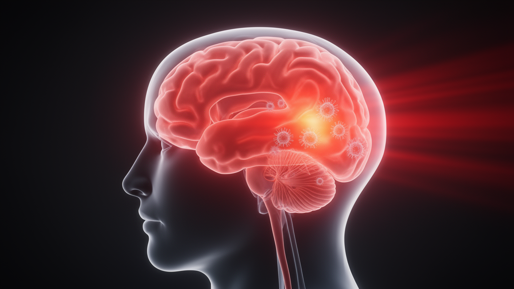
Brain disorders are now the leading cause of disability and the second leading cause of death worldwide, claiming more than 400 million disability-adjusted life years and roughly 11 million lives every year. Alzheimer’s and Parkinson’s diseases are climbing as populations age, while stroke, traumatic brain injury, and major depressive disorder cut across all age groups. Yet despite decades of research, most current treatments manage symptoms rather than the underlying disease mechanisms, and even the newest therapies often deliver only modest benefits or remain limited by accessibility and narrow therapeutic windows. Against this backdrop, a narrative review published in Frontiers in Neurology argues that a surprisingly simple intervention—low-intensity red to near-infrared light—may offer a way to target a process that unites these seemingly unrelated conditions.
That unifying process is neuroinflammation. Although it begins as a protective response of the central nervous system to injury, neuroinflammation can become chronic and maladaptive, amplifying damage across the entire spectrum of neurological disease. Microglial cells, the brain’s resident immune sentinels, are the main drivers of this response, with astrocytes playing a key modulatory role. When reactive microglia persistently release pro-inflammatory cytokines such as TNF-α, IL-1β, and IL-6, along with reactive oxygen species, they engage intracellular effectors like NF-κB and the NLRP3 inflammasome in a self-reinforcing loop that perpetuates inflammation. Chronic neuroinflammation is closely intertwined with oxidative stress, mitochondrial dysfunction, and disruption of the blood–brain barrier, which allows peripheral immune cells to infiltrate the brain and further fan the flames.
What makes neuroinflammation such an attractive therapeutic target is that whatever the initiating insult—amyloid-β and tau aggregates in Alzheimer’s disease, α-synuclein in Parkinson’s, autoimmune attack on myelin in multiple sclerosis, chronic stress in depression, or mechanical and ischemic injury in traumatic brain injury and stroke—these diverse triggers converge on a shared set of intracellular effectors. Redox imbalance, dysregulated nitric oxide, and altered calcium handling amplify cytokine production and sustain glial reactivity. Beyond the brain itself, systemic inflammation compromises the blood–brain barrier and sustains glial reactivity, and the brain–gut–microbiota axis adds another layer: gut dysbiosis has been implicated in Alzheimer’s, Parkinson’s, multiple sclerosis, stroke, and depression, modulating neuroinflammation through increased intestinal permeability, microbial metabolites, and vagal afferent pathways. Preclinical models even suggest the gut microbiota influences microglial maturation and their inflammatory phenotype.
Enter photobiomodulation, or PBM, formerly known as low-level laser therapy. The technique applies non-ionizing, non-thermal red to near-infrared light, typically in the 600 to 1,100 nanometer range, to biological tissues. When aimed at the brain, PBM is usually delivered transcranially, but photons are attenuated by overlying layers—hair, scalp, blood, and skull—before reaching brain tissue. PBM also follows a biphasic dose–response, in which low and high levels of light exert opposing effects, making wavelength selection, dosimetry, and anatomical targeting critical determinants of therapeutic efficacy. To overcome penetration limits, researchers have explored peripheral delivery strategies. In mouse and non-human primate models of Parkinson’s disease, PBM targeted at the abdomen or legs provided neuroprotection comparable to transcranial delivery, possibly reflecting an abscopal-like effect in which irradiated tissues release circulating mediators that modulate immune and metabolic responses at distant sites.
The mechanistic model that has emerged centers on mitochondrial photoreception. Photons are absorbed by cytochrome c oxidase, the terminal enzyme of the mitochondrial electron transport chain, enhancing mitochondrial activity and adenosine triphosphate production while modulating intracellular calcium, reactive oxygen species, nitric oxide availability, and cAMP-related signaling. Transient ROS modulation, together with activation of pathways such as ERK, PI3K/Akt, and Sirt1, influences transcription factors including NF-κB and Nrf2 that are central to inflammatory regulation. A growing body of evidence suggests cytochrome c oxidase is not the sole photoacceptor: heat- and light-gated transient receptor potential ion channels may contribute at specific wavelengths, and proposed roles for structured intracellular water remain hypothetical pending direct experimental validation. Clarifying these pathways is essential to refine wavelength- and dose-specific strategies.
At the cellular level, the most consistently reported target of PBM is microglial polarization. In vitro and in vivo studies show that PBM can shift reactive microglia from a pro-inflammatory profile—characterized by elevated iNOS, TNF-α, and IL-1β—toward a more regulatory state expressing Arg-1 and IL-10. Similar modulatory effects have been reported in astrocytes, where PBM inhibits neurotoxic polarization, preserves synaptic integrity, and promotes neuronal survival. In an APP/PS1 mouse model of Alzheimer’s disease, PBM reduced NLRP3 protein in both microglia and astrocytes, regulated inflammatory factor secretion, drove anti-inflammatory microglial polarization, and enhanced amyloid-β clearance. PBM has also been reported to attenuate NF-κB signaling in reactive microglia and macrophages, although the effect appears contact-dependent, with both inhibitory and stimulatory responses described depending on the baseline cellular activation state. In parallel, transient PBM-induced ROS modulation activates the Nrf2 pathway, upregulating endogenous antioxidant defenses.
Beyond direct effects on glial cells, PBM acts on mechanisms that contribute to structural and functional protection of neurons in an inflammatory context. Blood–brain barrier disruption, an early and shared feature of multiple neuroinflammatory conditions, has been shown to be attenuated by PBM through upregulation of tight junction proteins and restoration of barrier function via modulation of endothelial von Willebrand factor in a humanized hypoxic BBB model. PBM has also been associated with upregulation of brain-derived neurotrophic factor, supporting neuronal survival and synaptic plasticity. By coupling reduced inflammatory signaling with enhanced neurotrophic support, PBM may address both the injurious and reparative dimensions of neuroinflammatory pathology. Preclinical studies across disease models reinforce this multi-target signature: in Alzheimer’s models, transcranial and peripheral PBM reduced amyloid plaques and tau hyperphosphorylation, decreased microglial reactivity, restored mitochondrial function, and improved cognition, while combined brain–gut PBM normalized behavioral deficits and modulated gut microbiota composition. In Parkinson’s models, peripheral PBM conferred neuroprotection against MPTP-induced injury, preserving dopaminergic neurons in the substantia nigra with outcomes comparable to transcranial delivery. In experimental autoimmune encephalomyelitis, the standard model of multiple sclerosis, PBM reduced clinical severity, shifted spinal cytokine profiles toward a regulatory state, and prevented peripheral immune cell infiltration. In traumatic brain injury and stroke models, reduced secondary neuroinflammation was associated with improved cognitive and motor recovery, and in depression-related models, combined brain–gut PBM rescued cognitive impairments in chronically stressed mice alongside restoration of hippocampal Sirt1 levels and partial correction of stress-induced gut dysbiosis.
Clinical investigation, however, remains at an early stage, and the picture is decidedly mixed. In Alzheimer’s disease and mild cognitive impairment, cognitive benefits have been reported: a 2017 case series of five patients with mild-to-moderate dementia improved on MMSE and ADAS-Cog after 12 weeks of combined transcranial and intranasal PBM, though scores declined sharply during a four-week washout. A more recent investigation documented cognitive improvements alongside increased serum BDNF after 12 weeks of active transcranial PBM, and a randomized, double-blind, sham-controlled pilot trial of brain–gut PBM in 53 patients with mild-to-moderate Alzheimer’s demonstrated safety, high compliance, and trends toward cognitive improvement. Two ongoing trials—the TRAP-AD trial recruiting 196 patients to examine dose-dependent effects in early Alzheimer’s and amnestic MCI, and the multicentric Light4Life trial recruiting 108 patients for a six-month brain–gut intervention with cognitive, autonomy, and inflammatory biomarker outcomes—aim to provide more definitive answers. In Parkinson’s disease, by contrast, recent randomized sham-controlled trials have been largely negative on primary motor outcomes: a pilot study in 22 patients, a 12-week feasibility trial in 20 participants, and the largest sham-controlled study to date with 63 participants all failed to detect significant differences between active and sham groups on primary motor measures. In multiple sclerosis, a randomized trial reported increased serum IL-10 but no effect on nitrite levels, while a systematic review identified preliminary functional benefits. In stroke, the NEST program provides the largest clinical dataset: an initially positive phase II trial was followed by a phase III that did not confirm efficacy, and the pivotal NEST-3 trial with 630 patients was halted for futility, showing no difference from sham on 90-day functional outcome—a failure attributed in part to insufficient light delivery, a single treatment session, and the absence of dosimetric verification at the target tissue. In major depressive disorder, findings are the most discordant: a recent meta-analysis reported a moderate effect size for symptom reduction, yet the ELATED-3 multicentric sham-controlled trial observed no differences on its primary outcome among 49 subjects, concluding that a minimum dosimetric threshold is likely required, and a more recent trial demonstrated significant improvement in sleep quality but no difference on depression severity scales.
Several challenges must be resolved before PBM can claim a place in the neurological armamentarium. The foremost is dosimetric heterogeneity: wavelength, irradiance measurement, pulse frequency, and session duration and frequency vary considerably across studies, compounded by incomplete reporting of stimulation protocols, making cross-study comparison and dose–response characterization unreliable. Most studies reporting neuroprotective effects have used wavelengths in the 660 to 850 nanometer range, irradiances of roughly 10 to 100 milliwatts per square centimeter, and repeated sessions over several weeks. A further obstacle is the absence of validated biomarkers to confirm target engagement: most trials rely on cognitive and behavioral outcomes that offer little mechanistic insight into in vivo neuroinflammation modulation. Current options are limited—cerebrospinal fluid markers are invasive, and microglial PET tracers are expensive—though emerging blood-based biomarkers such as GFAP, and mechanistically interesting candidates like neuroglobin, a neuronal oxygen-binding globin that preserves mitochondrial function and limits free radical accumulation, may facilitate future assessment. Across all conditions, PBM has shown a consistently favorable safety profile with no serious adverse events reported, though long-term safety data remain limited. The review’s authors conclude that PBM may be best suited as an add-on rather than a standalone treatment, given that it acts on upstream pathways distinct from the symptomatic targets of current therapies—and that the possibility it is simply ineffective for certain indications, notably motor symptoms in Parkinson’s disease, must be entertained rather than attributed solely to suboptimal dosimetry. Continued efforts in mechanistic research, biomarker development, dosimetry standardization, and adequately powered clinical trials will determine whether light can truly become medicine for the inflamed brain.
Subject of Research: Photobiomodulation and neuroinflammation in brain disorders
Article Title: Photobiomodulation as a regulator of neuroinflammation: mechanistic insights and therapeutic potential in brain disorders
Article References: Blivet, G., Bertrand, C., Brauge, D., Delrieu, J., & Touchon, J. (2026). Photobiomodulation as a regulator of neuroinflammation: mechanistic insights and therapeutic potential in brain disorders. Frontiers in Neurology, 17, Article 1902491. https://doi.org/10.3389/fneur.2026.1902491
Image Credits: AI Generated
DOI: 10.3389/fneur.2026.1902491
Keywords: photobiomodulation, neuroinflammation, microglia, mitochondria, Alzheimer's disease, clinical trials, gut-brain axis, neurotechnology, regulator, mechanistic, insights, therapeutic
Cite Scienmag News
Cassandra Pierce. (October 17, 2026). Shining Light on the Brain: How Photobiomodulation Could Tame Neuroinflammation. Scienmag. https://scienmag.com/shining-light-on-the-brain-how-photobiomodulation-could-tame-neuroinflammation/
Cassandra Pierce. "Shining Light on the Brain: How Photobiomodulation Could Tame Neuroinflammation." Scienmag, 17 October 2026, https://scienmag.com/shining-light-on-the-brain-how-photobiomodulation-could-tame-neuroinflammation/. Accessed 17 October 2026.
Cassandra Pierce. "Shining Light on the Brain: How Photobiomodulation Could Tame Neuroinflammation." Scienmag. October 17, 2026. https://scienmag.com/shining-light-on-the-brain-how-photobiomodulation-could-tame-neuroinflammation/


