


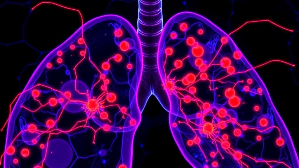
Lung adenocarcinoma, the most common form of lung cancer worldwide, has long been studied through the lens of its malignant cells: which mutations they carry, which drugs they resist, and how fast they divide. A new study published in Medical Oncology shifts that focus toward the geography of the tumor itself. By combining single-cell RNA sequencing, spatial transcriptomics, and large bulk transcriptomic cohorts from TCGA and GEO, a team led by Yixian Liang and Yong Li of the First Affiliated Hospital of Huzhou Normal University has mapped where lactate-glycolysis activity actually lives inside lung adenocarcinoma tumors, and the answer is not where many researchers might have expected. Rather than being confined to the cancer cells themselves, the most intense glycolytic transcriptional programs turned out to be concentrated in fibroblasts and neutrophils, the stromal and immune cells that surround and infiltrate the tumor.
The Warburg effect, the tendency of cancer cells to prefer glycolysis over oxidative phosphorylation even in the presence of oxygen, has been a cornerstone of tumor metabolism research for nearly a century. High glycolytic flux generates abundant lactate, which is exported into the tumor microenvironment, acidifying it and reshaping the behavior of neighboring immune cells. Previous work has shown that lactate can fuel regulatory T cells, suppress innate lymphoid cells, and promote immunosuppression across multiple cancer types. What has remained unclear, however, is the spatial architecture of this metabolic program in lung adenocarcinoma: which cells in the tumor actually run the glycolytic machinery, and how their locations relate to the immune landscape that determines whether a patient responds to immunotherapy.
To answer that question, the researchers needed a way to measure metabolic activity in individual cells while still knowing where those cells sit within the tumor. Their approach centered on AUCell-based scoring, a method that quantifies the activity of gene sets, in this case lactate-glycolysis-associated transcriptional programs, at single-cell resolution. Applied to single-cell RNA sequencing data from lung adenocarcinoma specimens, this scoring revealed a striking pattern: cells with high lactate-glycolysis activity were predominantly fibroblasts and neutrophils, not malignant epithelial cells. This finding challenges the assumption that tumor cells are the primary glycolytic engines of the tumor microenvironment and suggests that the metabolic burden of lung adenocarcinoma is shared, and perhaps even dominated, by its non-malignant residents.
The implications of that cellular distribution became clearer when the team examined how glycolytic activity related to immune infiltration and clinical outcomes. From the single-cell data, they distilled a compact metabolism-associated gene signature comprising three genes: RPS2, GAPDH, and LDHA. GAPDH, a canonical glycolytic enzyme, and LDHA, which converts pyruvate to lactate, are direct readouts of glycolytic flux, while RPS2, a ribosomal protein, reflects the translational activity that supports it. When projected onto bulk transcriptomic cohorts from TCGA and GEO, this three-gene signature was consistently correlated with immune remodeling and with an unfavorable prognosis. Patients whose tumors expressed higher levels of the signature fared worse, and their tumors showed distinct patterns of immune cell infiltration, pointing to a link between metabolic reprogramming and the composition of the immune microenvironment.
Correlation in bulk data, however, can be misleading. A gene signature elevated in aggressive tumors might simply reflect more cancer cells, more stress, or more necrosis rather than a genuine spatial relationship between metabolism and immunity. To resolve this, the investigators turned to spatial transcriptomics, a technology that preserves the physical coordinates of gene expression within intact tissue sections. The spatial data delivered the study’s most visually compelling result: GAPDH and LDHA expression co-localized with neutrophil-enriched regions of the tumor. In other words, the hottest glycolytic zones in lung adenocarcinoma are also the zones where neutrophils cluster, forming what the authors describe as a spatially structured, neutrophil-enriched immune remodeling pattern tied to lactate-glycolysis reprogramming.
Neutrophils are the most abundant white blood cells in the human body and are typically thought of as first responders to infection. In cancer, however, tumor-associated neutrophils have earned a more sinister reputation. Depending on signals from their microenvironment, they can adopt phenotypes that promote angiogenesis, suppress cytotoxic T cells, and facilitate metastasis, and high neutrophil-to-lymphocyte ratios are established markers of poor prognosis across many malignancies. The new findings suggest that lactate-rich, glycolytically active niches within lung tumors may be precisely the kind of environment that recruits or polarizes neutrophils, creating self-reinforcing neighborhoods where metabolism and immune suppression feed each other. Fibroblasts, the other major glycolytic cell type identified, are known architects of the tumor stroma, and their metabolic output could similarly condition the surrounding tissue.
Crucially, the team did not rely on computational analysis alone. They validated their key findings experimentally in clinical specimens using quantitative PCR and immunohistochemistry, confirming that the expression patterns of the signature genes observed in sequencing data hold up at the protein and transcript level in actual patient tissue. The research protocol was approved by the Medical Ethics Committee of Huzhou First People’s Hospital, and all participating patients provided written informed consent. This combination of multi-omics discovery and hands-on pathological validation gives the study a level of rigor that purely bioinformatic surveys often lack, and it anchors the abstract language of transcriptional programs in the physical reality of stained tissue sections.
The therapeutic implications are twofold. First, the three-gene signature of RPS2, GAPDH, and LDHA could serve as a stratification tool, helping clinicians identify lung adenocarcinoma patients whose tumors carry a metabolically driven, neutrophil-enriched immune configuration and therefore a worse outlook. Such patients might be candidates for more aggressive surveillance or for combination regimens that go beyond conventional immune checkpoint blockade. Second, the spatial coupling of glycolysis and neutrophil enrichment suggests that targeting lactate metabolism, for example through LDHA inhibition or lactate transport blockade, could disrupt the immunosuppressive niches that neutrophils inhabit, potentially converting a cold, hostile tumor microenvironment into one more receptive to immunotherapy. Metabolic inhibitors and immune modulators, the study implies, may need to be thought of together rather than separately.
There are, of course, important caveats. The study establishes associations between spatially resolved glycolytic programs and neutrophil-enriched immune remodeling; it does not yet prove that lactate production by fibroblasts and neutrophils causally drives neutrophil recruitment or immune suppression in lung adenocarcinoma. Functional experiments, such as disrupting glycolysis in specific stromal or immune compartments in model systems, would be needed to establish mechanism. The gene signature, while validated across cohorts and by experimental assays, will also require prospective clinical testing before it can inform patient care. Still, by demonstrating that lactate-glycolysis reprogramming in lung adenocarcinoma is spatially organized and intimately linked with a specific immune cell population, the work adds a crucial geographic dimension to cancer metabolism research. The tumor, this study reminds us, is not a uniform mass of hungry cancer cells but a patchwork of metabolic territories, and the boundaries of those territories may determine who lives and who dies.
Subject of Research: Spatially resolved lactate-glycolysis transcriptional programs and neutrophil-enriched immune remodeling in lung adenocarcinoma
Article Title: Spatially resolved lactate-glycolysis transcriptional programs are associated with neutrophil-enriched immune remodeling in lung adenocarcinoma
Article References: Liang, Y., Tan, J., Zhang, J., Wang, B., Shi, D., Ji, L., Li, X., Yu, D., & Li, Y. (2026). Spatially resolved lactate-glycolysis transcriptional programs are associated with neutrophil-enriched immune remodeling in lung adenocarcinoma. Medical Oncology, 43(10), Article 270. https://doi.org/10.1007/s12032-026-03390-8
Image Credits: AI Generated
DOI: 10.1007/s12032-026-03390-8
Keywords: lung adenocarcinoma, glycolysis, lactate metabolism, neutrophils, fibroblasts, spatial transcriptomics, single-cell RNA sequencing, tumor microenvironment, GAPDH, LDHA, cancer metabolism, prognosis
Cite Scienmag News
Nathaniel Bowman. (October 2, 2026). Lactate-Fueled Tumor Neighborhoods: Glycolysis Hotspots Recruit Neutrophils in Lung Cancer. Scienmag. https://scienmag.com/lactate-fueled-tumor-neighborhoods-glycolysis-hotspots-recruit-neutrophils-in-lung-cancer/
Nathaniel Bowman. "Lactate-Fueled Tumor Neighborhoods: Glycolysis Hotspots Recruit Neutrophils in Lung Cancer." Scienmag, 2 October 2026, https://scienmag.com/lactate-fueled-tumor-neighborhoods-glycolysis-hotspots-recruit-neutrophils-in-lung-cancer/. Accessed 4 October 2026.
Nathaniel Bowman. "Lactate-Fueled Tumor Neighborhoods: Glycolysis Hotspots Recruit Neutrophils in Lung Cancer." Scienmag. October 2, 2026. https://scienmag.com/lactate-fueled-tumor-neighborhoods-glycolysis-hotspots-recruit-neutrophils-in-lung-cancer/


