


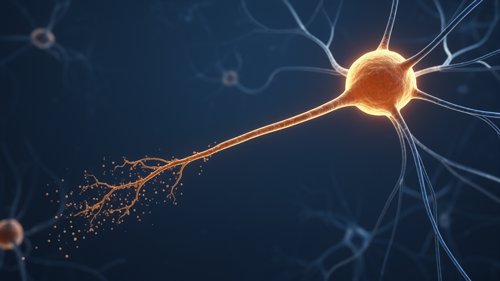
More than 160 years after Jean-Martin Charcot first described the hardened lateral columns of the spinal cord in patients dying of progressive motor deterioration, researchers have finally connected that hallmark lesion, known as lateral sclerosis, to the specific clinical signs it produces in people with amyotrophic lateral sclerosis. A new clinical-neuropathological study published in Acta Neuropathologica provides quantitative evidence that the degeneration of upper motor neurons in ALS behaves like a classic distal axonopathy: the longest, most distant reaches of the corticospinal tract fall apart first, and the damage then creeps back toward the neuron’s cell body in the motor cortex. The finding reframes a debate that has simmered in the field for decades about whether motor neuron disease spreads from the brain downward or from the periphery upward.
The research team, led by investigators at the University of Illinois at Chicago and the University of California, San Diego, took advantage of a rare and powerful resource: postmortem spinal cords from sporadic ALS patients whose clinical phenotypes had been carefully characterized during life by a single neurologist. Rather than studying a mixed patient population, the investigators deliberately selected the extremes of the clinical spectrum. Six patients had shown predominantly upper motor neuron signs at disease onset, including spasticity, hyperreflexia, pathological reflexes, and loss of fine skilled movements. Seven others had presented with predominantly lower motor neuron signs, marked by weakness, atrophy, and fasciculations. Thirteen neurologically healthy control spinal cords completed the comparison set.
The logic of this design is elegant. Upper motor neurons are the long projection neurons whose cell bodies sit in the motor cortex and whose axons descend through the brainstem, cross at the level of the pyramids, and form the lateral corticospinal tract, the great white-matter highway that carries commands to the lower motor neurons of the spinal cord. Because those axons are bundled together in a discrete tract, the lateral columns of the spinal cord offer the most direct window into upper motor neuron degeneration, something that is nearly impossible to assess by counting neurons in the cortex or by quantifying protein aggregates in brain tissue. If the clinical signs of upper motor neuron dysfunction truly reflect degeneration of this tract, then the tract should look profoundly damaged in patients with upper motor neuron-predominant disease and relatively spared in those with lower motor neuron-predominant disease.
That is precisely what the team found. Using Luxol Fast Blue-Periodic Acid Schiff staining to visualize myelin, the fatty insulation that wraps axons, the researchers documented severe, unmistakable myelin loss in the lateral corticospinal tract at all three spinal cord levels examined, cervical, thoracic, and lumbar, in every single one of the six upper motor neuron-predominant patients. In striking contrast, the lateral tracts of the lower motor neuron-predominant patients looked essentially indistinguishable from those of healthy controls at every level. The correlation between clinical phenotype and pathology followed an almost all-or-none pattern, providing the kind of clinicopathological validation that has been largely absent since Charcot’s era, when histological techniques were rudimentary by modern standards.
Myelin loss, however, was only the beginning of the story. The team developed a quantitative method to measure axon size distributions, generating frequency histograms of thousands of axons stained for neurofilament heavy chain, a core structural protein of the axonal skeleton. Previous studies had suggested that large-diameter axons, which originate from the giant Betz cells of the primary motor cortex and constitute the longest fibers in the corticospinal tract, are selectively lost in ALS. The new data extend that picture dramatically: axons of every caliber, from the smallest fibers measuring roughly 3.2 micrometers to the largest at about 16.4 micrometers, were significantly depleted in the upper motor neuron-predominant group, and the loss was consistently worse than in the lower motor neuron-predominant group at all three spinal levels. Sensory axons in the dorsal columns, examined as an internal comparison, were largely preserved, underscoring the specificity of the motor tract degeneration.
The most consequential observation emerged when the researchers compared axon loss between the cervical and lumbar segments of the cord. Because corticospinal axons travel from the brain downward, the lumbar cord represents the distal, far end of these extraordinarily long projections, while the cervical cord lies closer to their origin. In the upper motor neuron-predominant patients, axonal loss across all size ranges was significantly more pronounced in the lumbar cord than in the cervical cord, a rostrocaudal gradient that was absent in the lower motor neuron-predominant group. This length-dependent pattern is the anatomical signature of a dying-back process, the same phenomenon long recognized in peripheral neuropathies, where degeneration begins at the distant tips of long nerves and progresses proximally, producing the familiar stocking-glove pattern of sensory loss.
Woven through the degenerating tract, the researchers identified a striking inflammatory landscape. Foamy microglia, the brain’s resident immune cells swollen with lipid droplets after engulfing myelin and axonal debris, accumulated in large numbers precisely within the regions of most severe myelin and axon loss in the upper motor neuron-predominant cords. Quantification of Iba1-positive cells confirmed that these round, lipid-laden phagocytes were significantly increased at all three spinal levels compared with both the lower motor neuron-predominant group and controls. Notably, reactive astrocytes labeled by GFAP showed no comparable differences, and no foamy microglia were found in the sensory dorsal columns. The tight spatial coupling between foamy microgliosis and tract degeneration suggests an intimate glio-axonal interaction in upper motor neuron degeneration, although the study cannot determine whether these immune cells are drivers of the damage or simply the cleanup crew arriving after the collapse.
The study also delivered a surprise about TDP-43, the misfolded RNA-binding protein whose translocation from the nucleus to the cytoplasm is considered the pathological signature of ALS. While phosphorylated TDP-43 aggregates were rarely or never observed within the lateral corticospinal tract, a finding independently confirmed across three laboratories, total TDP-43 was measurably increased inside the axons of both the lateral and ventral corticospinal tracts when the protein was colocalized with phosphorylated neurofilament markers. Fibril-like axonal TDP-43 was also detected in the ventral horn gray matter, but the sensory axons of the dorsal columns showed no such elevation. This compartment-specific dysregulation hints that TDP-43 may behave differently in axons than in cell bodies, and differently in upper versus lower motor neurons, opening a new dimension for mechanistic studies.
The authors are careful to distinguish pattern from mechanism. Dying back, in their framing, is a descriptive term for distal-to-proximal degeneration, not a claim that sick axons are killing their neurons. Indeed, they argue, the most likely cause is a proximal failure within the neuron itself, probably in the cell body, that manifests first in the most distant axonal territory. Emerging evidence supports this view: when TDP-43 escapes the nucleus, it causes loss of function in transcripts such as STMN2, which maintains axon integrity, and UNC13A, which supports neuromuscular junction function, so a nuclear defect can silently starve the far reaches of an axon long before the cell body itself shows damage. The competing dying-forward hypothesis, which posits toxic antegrade spread from the motor cortex, remains speculative and has been criticized for its inability to explain the heterogeneity of ALS motor phenotypes.
The clinical implications are significant. Because upper motor neuron-predominant patients often survive longer, the extent of corticospinal tract destruction observed here was unexpectedly severe, suggesting a prolonged preclinical phase of slow but relentless degeneration. The findings also indicate that upper motor neuron degeneration progresses along two independent axes, one defined by the site of symptom onset and another by the relative balance of upper versus lower motor neuron involvement, with lumbar-projecting axons degenerating even in patients whose disease began in the bulbar region. For a field whose therapeutic pipeline has been dominated by neuron-centric strategies, this work makes a forceful case that the axon, its myelin sheath, and the foamy microglia that attend its demise deserve equal attention, and that length-dependent vulnerability may be a unifying principle across both tiers of the motor system in ALS.
Subject of Research: Distal axonopathy of upper motor neurons in amyotrophic lateral sclerosis
Article Title: Lateral sclerosis in amyotrophic lateral sclerosis is an upper motor neuron distal axonopathy (“dying back”): a clinical-neuropathological study
Article References: Liu, J., Ong, K., Meng, A., Stokin, G. B., Loeb, J. A., Ravits, J., & Song, F. (2026). Lateral sclerosis in amyotrophic lateral sclerosis is an upper motor neuron distal axonopathy (“dying back”): a clinical-neuropathological study. Acta Neuropathologica, 152(1), Article 48. https://doi.org/10.1007/s00401-026-03072-9
Image Credits: AI Generated
DOI: 10.1007/s00401-026-03072-9
Keywords: amyotrophic lateral sclerosis, lateral sclerosis, corticospinal tract, upper motor neuron, dying back axonopathy, myelin loss, TDP-43, foamy microglia, neuroinflammation, axonal degeneration, spinal cord pathology, motor neuron disease
Cite Scienmag News
Cassandra Pierce. (October 9, 2026). ALS’s ‘Lateral Sclerosis’ Revealed as a Dying-Back Degeneration of Upper Motor Neuron Axons. Scienmag. https://scienmag.com/alss-lateral-sclerosis-revealed-as-a-dying-back-degeneration-of-upper-motor-neuron-axons/
Cassandra Pierce. "ALS’s ‘Lateral Sclerosis’ Revealed as a Dying-Back Degeneration of Upper Motor Neuron Axons." Scienmag, 9 October 2026, https://scienmag.com/alss-lateral-sclerosis-revealed-as-a-dying-back-degeneration-of-upper-motor-neuron-axons/. Accessed 9 October 2026.
Cassandra Pierce. "ALS’s ‘Lateral Sclerosis’ Revealed as a Dying-Back Degeneration of Upper Motor Neuron Axons." Scienmag. October 9, 2026. https://scienmag.com/alss-lateral-sclerosis-revealed-as-a-dying-back-degeneration-of-upper-motor-neuron-axons/


