


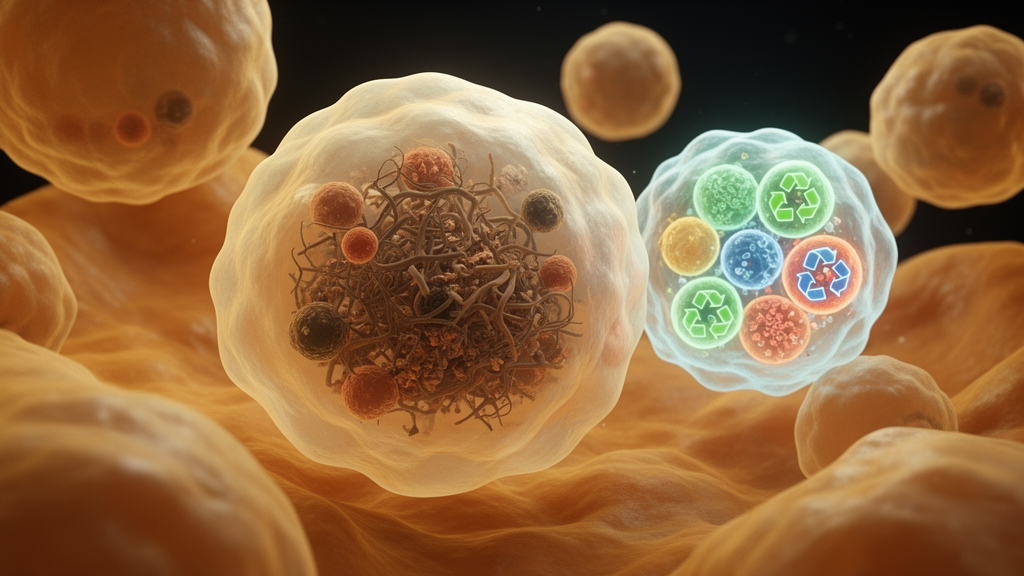
Inside every cell, a microscopic cleanup crew works around the clock, ferrying damaged and obsolete proteins to the lysosome for destruction. One of the most selective branches of this disposal system is chaperone-mediated autophagy, or CMA, a pathway that recognizes proteins bearing a specific molecular tag and shuttles them one by one through a channel in the lysosomal membrane. Now, a team led by Ana Maria Cuervo of the Albert Einstein College of Medicine has shown that the well-documented age-related decline of this pathway does far more than let cellular junk accumulate. Writing in Nature Aging, the researchers report that failing CMA fundamentally changes what senescent cells become and, crucially, whether the immune system can sweep them away. The findings reveal a vicious cycle at the heart of aging: worn-out recycling machinery makes senescent cells more toxic and simultaneously blinds the macrophages that are supposed to devour them.
CMA works with remarkable precision. The cytosolic chaperone HSC70 patrols the cell interior, binding proteins that carry a KFERQ-like amino acid motif and delivering them to LAMP2A, a receptor embedded in the lysosomal membrane. When substrates arrive, LAMP2A molecules multimerize into a translocation complex that unfolds and threads each protein into the lysosomal lumen for degradation. This process is not merely housekeeping. CMA regulates the cell cycle, metabolism, cellular differentiation, immune activation and even circadian rhythms. It also declines with age in most tissues, and its malfunction has been linked to neurodegenerative disease, metabolic syndrome and atherosclerosis. What remained unknown until now was how this decline shapes the fate of senescent cells, the growth-arrested cells that accumulate in aging organs and drive inflammation and tissue dysfunction.
The team began with mouse ear fibroblasts, a reliable primary model for studying aging outside the confounding effects of replicative senescence. Using fibroblasts from transgenic KFERQ-Dendra reporter mice, in which a fluorescent CMA substrate lights up as puncta inside lysosomes, the researchers tracked CMA activity as cells entered senescence. In fibroblasts from young, four-month-old mice, senescence induction with the CDK4/6 inhibitor palbociclib triggered a robust upregulation of CMA, consistent with earlier work showing that the pathway is activated when cells remodel their proteome. Fibroblasts from twenty-three-month-old mice, however, told a different story. They displayed lower basal CMA activity and completely failed to boost the pathway in response to the senescence stimulus. The deficit was not due to reduced reporter expression but to impaired delivery and internalization of substrates into lysosomes, confirming that aged cells lose the ability to engage CMA precisely when they need it most.
To dissect the consequences, the researchers stably knocked down Lamp2a, the rate-limiting component of CMA, in untransformed NIH3T3 fibroblasts. Strikingly, these CMA-deficient cells acquired several senescence-like features even before any senescence stimulus: elevated senescence-associated beta-galactosidase staining, nuclear accumulation of the chromatin protein HMGB1, partial increases in p21 and the DNA damage marker gamma-H2A.X, and nuclear indentations. Yet they kept proliferating normally and arrested only after palbociclib treatment, indicating that CMA loss alone does not drive full conventional senescence. Principal component analysis of multiple readouts placed the knockdown cells in an intermediate cluster between healthy controls and senescent cells. The message was clear: CMA is dispensable for initiating senescence, but its loss warps the senescence program itself, producing cells with properties that resemble those of aged senescent cells.
Quantitative proteomics revealed just how deep that resemblance runs. Blocking CMA in young cells reproduced nearly half of the proteome changes seen in aged cells, with shared alterations concentrated in lipid metabolism, oxidative phosphorylation, protein translation, vesicular trafficking and extracellular matrix remodeling. When young control, young CMA-deficient and aged fibroblasts were all pushed into senescence, the CMA-deficient and aged groups showed forty-five percent concordance in their proteome remodeling, with close to four hundred proteins changing in parallel and only about two hundred differing between them despite their twenty-month age gap. By inhibiting lysosomal proteolysis and comparing degradation profiles, the team identified sixty-four proteins that are no longer degraded in lysosomes in aged cells, more than half of them bona fide CMA substrates. In other words, a substantial fraction of the aging proteome is simply a backlog of proteins that the failing CMA machinery can no longer clear.
Metabolomics added a second layer of evidence. Under basal conditions, CMA-deficient fibroblasts phenocopied many metabolic features of aged cells, including changes in energy production, carbohydrate and glycosylation pathways, lipid and membrane biosynthesis, and amino acid and methylation pathways. Upon senescence induction, young control cells mounted a characteristic metabolic shift, engaging fatty acid beta-oxidation, while CMA-deficient and aged cells failed to make that transition and instead showed unique changes in the tricarboxylic acid cycle, aromatic amino acid metabolism and nucleotide metabolism. An integrative analysis combining proteomic and metabolomic datasets confirmed that proteins normally degraded by CMA during senescence accounted for a large share of these metabolic differences, particularly the failure to engage fatty acid and one-carbon metabolism. Because the senescence-associated secretory phenotype, or SASP, depends on this metabolic reprogramming, the stage was set for a dramatic effect on what senescent cells secrete.
That effect materialized in the secretome. Using an ER-TurboID system that biotinylates proteins inside the endoplasmic reticulum, the researchers separated conventionally secreted proteins from those released through unconventional routes such as exosomes and lysosomal docking. CMA-deficient cells showed a deregulated basal secretome and a profoundly altered response to senescence: rather than completing a normal SASP, they failed to downregulate thirty-nine conventionally secreted proteins and upregulated forty-seven others involved in cell migration, protein folding and lysosomal enzymes, with pronounced enrichment of immune-related factors. Unconventionally secreted nuclear proteins appeared in the medium, a selective release not attributable to cell death. Functionally, the medium from CMA-deficient cells, even before senescence induction, drove paracrine senescence in healthy neighboring fibroblasts, raising SA-beta-gal positivity, p21 levels and nuclear HMGB1 loss to degrees comparable to medium from senescent control cells. The secreted metabolites were similarly skewed toward proinflammatory signaling and known regulators of macrophage function.
The most consequential discovery concerned macrophages, the professional scavengers that clear senescent cells through phagocytosis and efferocytosis. Bone marrow-derived macrophages from aged mice showed significantly reduced CMA activity, and exposure to the secretome of senescent fibroblasts suppressed CMA in young macrophages, with the strongest inhibition from CMA-deficient senescent cells. Macrophages engineered to lack LAMP2A displayed reduced phagocytosis and impaired efferocytosis of dying cells, and co-culture experiments showed senescent fibroblasts persisting in their presence. The mechanism traced to SIRPalpha, a phagocytosis-inhibitory receptor that is normally degraded via CMA after endocytic internalization. Without CMA, SIRPalpha accumulates on the macrophage surface, recruits the phosphatase SHP-1, and amplifies the CD47-SIRPalpha ‘do not eat me’ signal, suppressing the actin remodeling that engulfment requires. Aged macrophages showed the same elevated SIRPalpha and SHP-1 levels. In mice lacking LAMP2A specifically in macrophages, senescent cell burden rose in adipose tissue, liver, lung and brain, and full-thickness wounds healed more slowly, with lingering p21-positive cells despite abundant macrophages at the injury site.
The therapeutic implications were tested with CA77.1, a small-molecule CMA activator. Oral administration to eighteen-month-old mice for five months prevented the age-related rise in senescent cells across adipose tissue, liver and lung, dampened inflammatory and senescence gene expression, reduced fibrosis, and restored the phagocytic activity of aged macrophages to levels comparable to those of young animals. In a bleomycin model of pulmonary fibrosis, early CMA activation preserved body weight, cut lung fibrosis, lowered collagen and p16 expression, and normalized the expansion of lung macrophages, while late treatment was far less effective. Human data lent clinical weight: a transcriptional CMA score declined with age in lung cells and was significantly reduced in patients with idiopathic pulmonary fibrosis, whose isolated lysosomes showed unstable LAMP2 and reduced substrate uptake. Together, the results reframe senescent cell accumulation not as a failure of senescence itself but as a failure of its resolution, and they position CMA upregulation as a dual-action strategy, simultaneously improving the health of senescent cells’ secretions and re-arming the immune cells that must dispose of them.
Subject of Research: How age-related decline of chaperone-mediated autophagy alters senescent cells and impairs macrophage-mediated immune clearance
Article Title: Decline of chaperone-mediated autophagy in aging impairs macrophage clearance of senescent cells
Article References: Sereda, R., Lindenau, K., Diaz, A., Liu, Z., Santiago-Fernández, O., Khawaja, R. R., Cutler, R., Calyeca, J., McCabe, M., Durand, S., Aprahamian, F., Vanegas, N. D. P., Chen, H., Vilicich, F., Chavda, B., Botbol, Y., Kroemer, G., Finkel, T., Gavathiotis, E., … Cuervo, A. M. (2026). Decline of chaperone-mediated autophagy in aging impairs macrophage clearance of senescent cells. Nature Aging. https://doi.org/10.1038/s43587-026-01240-w
Image Credits: AI Generated
DOI: 10.1038/s43587-026-01240-w
Keywords: chaperone-mediated autophagy, cellular senescence, macrophages, aging, LAMP2A, SASP, efferocytosis, proteomics, metabolomics, pulmonary fibrosis, CA77.1, lysosomes
Cite Scienmag News
Beatrice Stafford. (October 9, 2026). Aging Cells Lose Their Recycling Machinery, Letting Senescent Cells Pile Up. Scienmag. https://scienmag.com/aging-cells-lose-their-recycling-machinery-letting-senescent-cells-pile-up/
Beatrice Stafford. "Aging Cells Lose Their Recycling Machinery, Letting Senescent Cells Pile Up." Scienmag, 9 October 2026, https://scienmag.com/aging-cells-lose-their-recycling-machinery-letting-senescent-cells-pile-up/. Accessed 9 October 2026.
Beatrice Stafford. "Aging Cells Lose Their Recycling Machinery, Letting Senescent Cells Pile Up." Scienmag. October 9, 2026. https://scienmag.com/aging-cells-lose-their-recycling-machinery-letting-senescent-cells-pile-up/


