


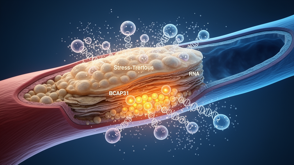
Atherosclerotic plaques are not inert deposits of fat. They are chronically inflamed tissues in which macrophages accumulate, die, and fail to be cleared, and in which the balance between cell survival and cell loss helps determine whether a lesion stays stable or becomes rupture-prone. A new integrative study published in Frontiers in Cell Development and Biology combines four independently phenotyped human carotid plaque transcriptomic datasets with laboratory perturbation experiments to argue that one endoplasmic reticulum membrane protein, BCAP31, behaves less like a disease driver and more like a cellular stress buffer in plaque macrophages.
The research team, led by investigators at Wenzhou Central Hospital in China, began with paired bulk transcriptomes from 32 patients, each contributing both a carotid plaque sample and a sample of macroscopically intact adjacent tissue. Using linear models that accounted for patient identity, they scored seven prespecified gene programs: apoptosis, ferroptosis, pyroptosis, necroptosis, inflammasome signaling, the general inflammatory response, and TNF signaling through NF-κB. All seven rose in plaque tissue, with false-discovery rates ranging from roughly 10⁻⁷ to 10⁻⁵. Because the gene sets overlap only minimally, the authors argue that this coordinated increase reflects genuinely parallel activation of multiple death and inflammatory programs rather than an artifact of duplicated gene lists.
To place these tissue-level signals in clinically meaningful contexts, the team turned to two instability cohorts. Laser-captured macrophages from five ruptured and six stable plaques showed elevated ferroptosis, pyroptosis, and necroptosis scores in the ruptured group, while apoptosis and the inflammatory programs did not survive correction. Whole-plaque transcriptomes from 27 lesions with intraplaque hemorrhage and 16 without showed a broader pattern: all seven programs were significantly increased in hemorrhagic plaques. The two instability phenotypes thus carried overlapping but non-identical death-associated profiles, underscoring that plaque vulnerability is not a single molecular state.
BCAP31, which encodes the B-cell receptor-associated protein 31, emerged from a cross-cohort screen. The gene was increased in paired plaque tissue with a log2 fold change of 0.375, in intraplaque-hemorrhage plaque with a log2 fold change of 0.901, and in ruptured macrophages with a log2 fold change of 0.972, although the rupture result was only nominally significant and did not survive transcriptome-wide correction. In a weighted coexpression network built from the discovery cohort, BCAP31 belonged to a 1,999-gene module whose eigengene increased in plaque, and the module was enriched for immune and cytokine-related biological processes.
Single-cell RNA sequencing from three patients with matched atherosclerotic core and proximal adjacent tissue allowed the team to ask where the BCAP31 signal resided. After stringent quality control reduced 51,275 input cells to 41,528 singlets, marker-audited clustering resolved twelve cell classes. Patient-level pseudobulk aggregation, in which counts were summed per patient, condition, and cell type before statistical testing, showed that BCAP31 rose in macrophages with a log2 fold change of 0.756, and the direction was positive in all three patients. Importantly, this estimate did not remain significant after correction across all 49 cell-type and program combinations, so the authors treated it as a localization signal rather than a transcriptome-wide significant single-cell association.
The macrophage compartment itself proved heterogeneous. Reclustering resolved five states, including inflammatory monocytes, HLA-C1QC-expressing macrophages, APOC1-SPP1 remodeling macrophages, and SPP1-expressing lipid-loaded macrophages. Compared with adjacent tissue, plaque core samples showed increases in the two SPP1-rich remodeling states and a decrease in inflammatory monocytes, each directionally consistent across all three patients. Cross-sectional ordering analyses placed higher BCAP31 expression toward the plaque-enriched remodeling endpoints, and predicted communication modeling suggested that macrophage-to-vascular-wall SPP1 signaling increased in all three patient pairs. The authors are careful to note that these ordering and communication outputs are model-based descriptions of transcriptional placement, not evidence of temporal progression or measured ligand binding.
The decisive question was what BCAP31 actually does when its abundance changes. In THP-1-derived macrophages exposed to oxidized low-density lipoprotein, the team silenced BCAP31 with siRNA and measured the consequences. Depletion amplified the oxLDL-induced expression of the ER-stress transcripts HSPA5, ATF4, DDIT3, and spliced XBP1, and increased nuclear accumulation of the DDIT3 protein, a key effector of ER-stress-associated apoptosis. Inflammatory output rose in parallel: transcripts and secreted protein levels of TNF-alpha, IL-1beta, and IL-6 all increased, and Annexin V/propidium iodide flow cytometry revealed more apoptotic cells in the depleted, oxLDL-exposed group. Notably, depletion also raised some ER-stress readouts even without oxLDL, hinting at a basal role for BCAP31 in maintaining ER homeostasis.
To test whether ER stress was mechanistically linked to this phenotype, the researchers added 4-phenylbutyrate, a chemical chaperone that eases protein folding burden in the ER. The compound lowered the ER-stress transcripts, reduced cytokine release, and decreased Annexin V-positive apoptosis in the BCAP31-depleted, oxLDL-exposed cells. The concordant attenuation across three assay types supports ER-stress involvement in the phenotype. The authors caution, however, that because the design lacked an oxLDL-plus-chaperone group with intact BCAP31, the experiments cannot separate general pharmacological protection from a BCAP31-specific reversal of the phenotype.
The direction of these perturbation results is what gives the study its interpretive punch. If BCAP31 upregulation were simply pathogenic, removing it should have protected the cells. Instead, removing it made things worse. The authors therefore propose that the elevated BCAP31 seen in vulnerable plaque contexts reflects a compensatory, stress-adaptive response: macrophages facing oxidized lipids and proteotoxic pressure increase BCAP31 transcripts in an attempt to preserve ER function, and the elevation marks greater demand for proteostasis rather than a cause of injury. This reading also fits BAP31’s known dual molecular behavior. Full-length, uncleaved BAP31 can restrain Fas-associated mitochondrial apoptosis, whereas caspase cleavage generates a p20 fragment that promotes ER calcium release, mitochondrial fission, and cytochrome-c mobilization. Because the study measured mRNA and not protein cleavage states, it cannot resolve which molecular form predominates in advanced lesions.
The limitations are clearly stated and worth keeping in view. The single-cell cohort contained only three patients and the rupture cohort only eleven, limiting statistical power and generalizability. Whole-plaque associations may partly reflect shifts in cellular composition rather than within-cell changes, and PMA-differentiated THP-1 cells do not fully reproduce the ontogeny or multicellular environment of primary plaque macrophages. Even so, the study illustrates a methodological lesson for atherosclerosis research: tissue-level enrichment of death programs cannot be assigned wholesale to every lineage, as shown by the finding that fibroblast apoptosis actually decreased in plaque while macrophage apoptosis scores did not change. By coupling patient-matched human transcriptomics with directional perturbation experiments, the work moves a candidate gene beyond association and toward a mechanistic hypothesis, one that positions BCAP31 as a marker, and possibly a mediator, of the stress-adaptive capacity of plaque macrophages.
Subject of Research: The role of BCAP31 in endoplasmic reticulum stress, inflammation, and apoptosis in human carotid atherosclerotic plaque macrophages
Article Title: Integrated human carotid plaque transcriptomics and macrophage perturbation link BCAP31 to endoplasmic reticulum stress, inflammation, and apoptosis
Article References: Wang, Y., Lin, B., Chen, S., Yang, J., & Huang, A. (2026). Integrated human carotid plaque transcriptomics and macrophage perturbation link BCAP31 to endoplasmic reticulum stress, inflammation, and apoptosis. Frontiers in Cell and Developmental Biology, 14, Article 1970662. https://doi.org/10.3389/fcell.2026.1970662
Image Credits: AI Generated
DOI: 10.3389/fcell.2026.1970662
Keywords: BCAP31, atherosclerosis, carotid plaque, macrophages, endoplasmic reticulum stress, apoptosis, inflammation, intraplaque hemorrhage, plaque rupture, transcriptomics, single-cell RNA sequencing, oxidized LDL
Cite Scienmag News
Brooke Gardner. (October 23, 2026). Plaque Transcriptomics Point to BCAP31 as a Stress Buffer in Carotid Atherosclerosis. Scienmag. https://scienmag.com/plaque-transcriptomics-point-to-bcap31-as-a-stress-buffer-in-carotid-atherosclerosis/
Brooke Gardner. "Plaque Transcriptomics Point to BCAP31 as a Stress Buffer in Carotid Atherosclerosis." Scienmag, 23 October 2026, https://scienmag.com/plaque-transcriptomics-point-to-bcap31-as-a-stress-buffer-in-carotid-atherosclerosis/. Accessed 23 October 2026.
Brooke Gardner. "Plaque Transcriptomics Point to BCAP31 as a Stress Buffer in Carotid Atherosclerosis." Scienmag. October 23, 2026. https://scienmag.com/plaque-transcriptomics-point-to-bcap31-as-a-stress-buffer-in-carotid-atherosclerosis/


