


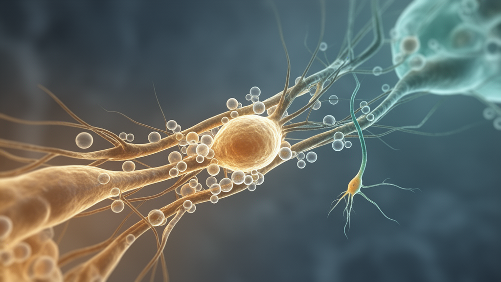
A 2026 mini review concludes that changes in microglial energy metabolism may help connect pathological conditions in the brain with remodeling of the niches in which adult neurons are generated. Drawing on evidence from animals, cultured cells and human postmortem tissue, the authors identify immunometabolism as a possible interface between microglial activity and the surrounding environment of neural stem cells. Metabolic state is associated with phagocytosis, inflammatory signaling, synaptic pruning, neurotrophic support and extracellular-vesicle communication, all of which can affect neurogenesis.
The evidence does not establish one metabolic pathway or a universal microglial state as the cause of neurogenic change, however, and many of the proposed links remain based on associations or experimental systems that did not measure adult neurogenesis directly. Adult neurogenesis involves the activation and differentiation of neural stem cells, followed by the survival and integration of newly generated neurons into circuits associated with memory and emotional regulation. In mammals, the process persists in the subventricular and subgranular zones, although its contribution to human brain homeostasis remains debated.
The neurogenic niche is a multicellular microenvironment containing neural stem cells, neurons, vasculature and glia that regulates stem-cell quiescence, fate specification and survival. Because these surrounding cells provide signals and conditions that support or constrain stem-cell activity, impaired neurogenesis can reflect changes in the niche as well as alterations intrinsic to neural stem cells. Microglia are resident immune cells that act as active participants in this environment.
Under physiological conditions, they survey surrounding tissue, recognize and engulf apoptotic cells and debris, prune synapses and provide neurotrophic signals. These tasks require substantial bioenergetic capacity, particularly for continuous surveillance, migration and phagocytosis. The review notes that microglia predominantly rely on mitochondrial oxidative phosphorylation to meet these demands.
Pathological stimulation can instead produce metabolic reprogramming: increased glycolysis is generally associated with pro-inflammatory responses, while changes in mitochondrial activity, reactive oxygen species production, glucose metabolism, lipid storage and metabolic flexibility can accompany shifts in cellular function. These relationships are not fixed, because the same metabolic feature can be associated with different microglial states depending on the stimulus and disease context. Depression and aging illustrate two forms of neurogenic niche dysregulation described in the review.
Chronic stress suppresses hippocampal neurogenesis, and rodent studies have associated this response with reduced brain-derived neurotrophic factor and insulin-like growth factor 1 expression, elevated inflammatory cytokines and altered microglial markers. Transcriptomic analysis of postmortem brain tissue from people with major depressive disorder identified an immunosuppressive microglial phenotype, including differential regulation of pathways involved in complement signaling and phagocytosis. In a mouse model of chronic stress, electroacupuncture promoted BDNF-dependent neurogenesis alongside microglial reprogramming.
In postpartum C57BL/6J dams exposed to pup separation and chronic restraint stress, reduced hippocampal inflammation and microglial activation were associated with increased adult hippocampal neurogenesis and stress resilience. The authors caution that direct evidence linking postpartum endocrine and immune changes to microglial metabolism remains limited. Aging is associated with reduced neural stem-cell activation and diminished production of newborn neurons, together with progressive impairment of microglial surveillance, migration and phagocytic functions.
Senescent microglia can exhibit mitochondrial dysfunction, reduced ATP production, accumulation of reactive oxygen species, abnormal glucose metabolism, lipid-droplet accumulation and decreased metabolic flexibility. The resulting inflammatory changes and loss of homeostatic activity can overlap rather than forming entirely separate patterns. In rats with hippocampal IL-1β overexpression, microglial lipid droplets increased while adult hippocampal neurogenesis decreased.
This association is consistent with a metabolic contribution, but it does not show that lipid accumulation caused the neurogenic deficit. More broadly, age-related neurogenic decline coincides with, and may partly reflect, weakening of the niche support supplied by microglia. Phagocytosis offers one of the clearest connections between microglial function and adult neurogenesis.
During neurogenesis, a substantial proportion of newborn cells undergoes programmed cell death at an early developmental stage, making prompt clearance of apoptotic cells important for niche homeostasis. Microglia rapidly engulf this material, helping prevent persistent local inflammation. Engulfment also initiates a transcriptional program that changes the microglial secretome and can influence adult hippocampal neurogenesis.
In mice subjected to social defeat stress, microglial engulfment of neural stem cells increased while hippocampal neurogenesis was impaired. Neural stem-cell-specific deletion of TRPV4 and pharmacological removal of microglia each ameliorated the stress-associated neurogenic impairment in that model. In separate experiments, mice deficient in P2Y12 or in MerTK/Axl signaling showed reduced microglial phagocytosis, lower adult hippocampal neurogenesis and reduced neuronal production.
Together, these findings show that controlled phagocytic activity belongs to normal niche regulation, while excessive or altered engulfment can accompany neurogenic impairment. Microglia also participate in the synaptic reorganization needed for newborn neurons to enter existing neural networks. Complement proteins can tag particular synapses for elimination, and complement C3/CR3 activation has been associated with aberrant microglia-mediated synaptic pruning and reduced expression of synaptic proteins in a mouse model of chronic unpredictable mild stress.
Because complement signaling is itself involved in neurogenesis, altered pruning may connect neural production with circuit integration. Other microglial outputs are trophic and extracellular. In lupus-prone mice with hippocampal dysfunction, microglial activation and increased pro-inflammatory cytokines accompanied disrupted hippocampal neurogenesis and greater apoptosis of adult neural stem cells.
Mitochondrial dysfunction has been associated with reduced IGF-1 and BDNF expression, while promoting mitochondrial biogenesis can enhance markers of reparative microglial states and increase BDNF secretion. Microglia-derived extracellular vesicles add another communication route, carrying nucleic acids, proteins and metabolism-related molecules that can affect neural stem-cell fate, glial crosstalk and local signaling. The cargo of these vesicles can change under pathological conditions.
In a binge-ethanol exposure model, pro-inflammatory microglial extracellular vesicles carried increased G9a mRNA, raised G9a/H3K9me2 levels in recipient cells and impaired adult hippocampal neurogenesis. Separate work in an Alzheimer’s disease model found that extracellular vesicles derived from human induced pluripotent stem-cell-derived neural stem cells were internalized by microglia and neurons and contacted astrocytes. Treatment with those vesicles reduced inflammatory signaling in microglia and astrocytes and improved cognitive and mood measures.
This result shows that intercellular communication can connect microglia with other cells, but it does not establish that a particular microglial metabolic program caused the treatment effect. The review also cites work in which microglia-derived exosomes delivered pro-neurogenic microRNAs that promoted neuronal differentiation of neural stem cells and neural regeneration at injury sites, providing experimental evidence for extracellular-vesicle signaling without establishing a clinical intervention. Pathological mitochondrial changes may reshape the niche environment more broadly.
In an ischemic stroke model, inhibiting JAK2/STAT3 signaling reduced Drp1 phosphorylation, mitochondrial translocation and fission in microglia, accompanied by reduced neuroinflammation. Drp1 is a key regulator of mitochondrial fission and is closely associated with inflammatory responses. Mitochondrial complex I reverse electron transport can accelerate reactive oxygen species generation, while excessive oxidative stress can inhibit neural stem-cell proliferation and differentiation.
In an Alzheimer’s disease-related experimental model, amyloid-beta exposure induced oxidative stress, endoplasmic-reticulum stress and mitochondrial dysfunction in human neural stem cells, including impaired ATP production and reduced mitochondrial respiration. Metabolites such as lactate, succinate and ketone bodies may also act as signals that modify redox states, epigenetic regulation and cellular pathways. Metabolomic studies have identified reduced glycolytic and Krebs-cycle markers, increased ketone bodies and amino-acid catabolism-related metabolites, and increased phosphatidylcholine 40:2 in Parkinson’s disease, but direct evidence that metabolites released by microglia control adult neurogenesis remains limited.
The review identifies possible intervention points involving mitochondrial biogenesis, mitochondrial dynamics, clearance of damaged mitochondria and metabolic-sensing pathways such as AMPK, mTOR, SIRT1 and PGC-1α. Extracellular-vesicle transfer is another proposed route for reshaping communication between microglia and neural stem cells. These strategies remain preclinical: findings come from cultured cells, rodents, ex vivo models or disease-specific experimental systems, and some experiments did not directly assess adult neurogenesis.
For example, targeting microglial lipid metabolism in obesity increased phagocytosis, reduced inflammation and improved metabolic flexibility, but neurogenesis was not measured. Mitochondrial transplantation is another experimental approach; microglia internalized exogenous mitochondria, with reported increases in ATP and reduced reactive oxygen species accumulation, while its contribution to neurogenesis was not established. The authors’ central conclusion is that microglial immunometabolism deserves study as a possible link between pathological disturbances in the brain and localized neurogenic decline.
Subject of Research: Microglial immunometabolism in adult neurogenic niche remodeling
Article Title: Microglia in the neurogenic niche: emerging roles of immunometabolism
Article References: Liu, D., Li, Y., & Liu, Y. (2026). Microglia in the neurogenic niche: emerging roles of immunometabolism. Frontiers in Neuroscience, 20, Article 2007175. https://doi.org/10.3389/fnins.2026.2007175
Image Credits: AI Generated
DOI: 10.3389/fnins.2026.2007175
Keywords: microglia, adult neurogenesis, immunometabolism, neurogenic niche, neural stem cells, phagocytosis, mitochondrial dysfunction, synaptic pruning, extracellular vesicles, brain aging, depression, neurogenic
Cite Scienmag News
Daisy Hatcher. (October 22, 2026). Review links microglial metabolism to adult neurogenesis regulation. Scienmag. https://scienmag.com/review-links-microglial-metabolism-to-adult-neurogenesis-regulation/
Daisy Hatcher. "Review links microglial metabolism to adult neurogenesis regulation." Scienmag, 22 October 2026, https://scienmag.com/review-links-microglial-metabolism-to-adult-neurogenesis-regulation/. Accessed 22 October 2026.
Daisy Hatcher. "Review links microglial metabolism to adult neurogenesis regulation." Scienmag. October 22, 2026. https://scienmag.com/review-links-microglial-metabolism-to-adult-neurogenesis-regulation/


