


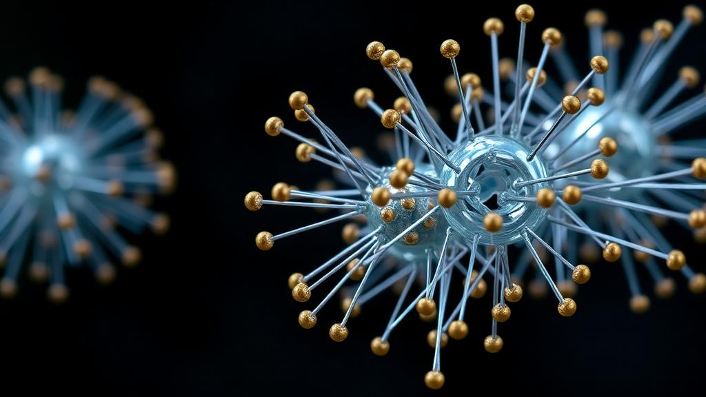
Inorganic nanoparticles, particles engineered at scales between one and one hundred nanometers, are rapidly becoming one of the most consequential tools in modern oncology. A new narrative review published in the journal Pharmacia synthesizes five years of research on these materials as drug delivery systems, arguing that their unique magnetic, optical, and catalytic properties position them at the heart of theranostics, the increasingly clinical practice of merging diagnosis and therapy into a single personalized intervention. Drawing on peer-reviewed studies published between 2020 and 2025 and indexed in PubMed, Scopus, and Web of Science, the review maps both the extraordinary promise of these platforms and the unresolved toxicological questions that still stand between the laboratory and the clinic.
The appeal of inorganic nanoparticles over their organic counterparts is rooted in physics. Their high surface area-to-volume ratio, tunable particle size, enhanced stability, and ease of surface functionalization allow researchers to design carriers that are simultaneously stable in the bloodstream, efficient at loading drugs, and responsive to external triggers. Unlike conventional chemotherapy, which floods the body with cytotoxic compounds and is limited by dose-limiting toxicities such as cardiotoxicity and neutropenia, nanoparticle-based systems can concentrate therapeutic payloads at tumor sites while sparing healthy tissue. A recent meta-analysis cited in the review found that nanoparticle-based chemotherapy delivered significantly improved overall survival, progression-free survival, and objective response rates compared with conventional regimens, alongside lower treatment-related toxicity.
Among the most transformative platforms are magnetic nanoparticles, which can be steered and activated by alternating magnetic fields. These carriers combine three functions in one system: targeted drug delivery, magnetic hyperthermia therapy in which the field raises the temperature at the tumor site and kills cancer cells, and magnetic resonance imaging contrast for real-time visualization. Magnetite nanoparticles, with a mass-specific magnetization of roughly 92 emu per gram, retain magnetization even without an external field, making them exceptionally navigable, while maghemite offers somewhat lower magnetization at around 74 emu per gram but better oxidative stability and lower toxicity. Cobalt ferrite, nickel ferrite, iron-platinum alloys, and rare-earth iron-boron compounds round out a family of materials whose magnetic properties can be matched to specific clinical tasks.
The most striking application is in neuro-oncology. The blood-brain barrier, a selective cellular gatekeeper, blocks most chemotherapeutic and immunotherapeutic drugs from reaching brain tumors, and the infiltrative, heterogeneous nature of gliomas frustrates surgery. Magnetic nanoparticles, particularly superparamagnetic iron oxide nanoparticles, have repeatedly demonstrated the ability to cross this barrier while minimizing systemic toxicity. The review distinguishes three targeting strategies: passive targeting that exploits the enhanced permeability and retention effect, in which particles between roughly 3 and 200 nanometers leak into tumor capillaries and become trapped; active targeting, in which ligands such as transferrin, folate, hyaluronic acid, aptamers, antibodies, and peptides are conjugated to the particle surface to bind receptors overexpressed on cancer cells; and magnetic targeting itself, in which an externally positioned magnetic field physically drags drug-laden particles to the tumor. Notably, several studies report that magnetic targeting outperforms both passive and ligand-mediated approaches in brain tumor models.
A second frontier involves metal-organic frameworks, porous nanocomposites that marry metal ions with organic ligands to create structures of enormous surface area. Porphyrin-based frameworks act simultaneously as drug carriers and photosensitizers, generating reactive oxygen species that penetrate cancer cells and drive apoptosis. One recently described nanocomposite, PCN-222/Au/DOX, combines a rod-shaped porphyrinic framework with gold nanoparticles and a low dose of doxorubicin, producing multifunctional anticancer and antibacterial activity against Helicobacter pylori and highlighting its potential in gastric cancer. A separate guanidinium-based framework, GZHMU-1@Mo, catalyzes the production of reactive oxygen species from oxygen and hydrogen peroxide inside tumor cells while degrading their antioxidant reserves, disrupting the intracellular redox balance in an approach known as chemodynamic therapy, which offers deep-tissue treatment without drug resistance.
The review also highlights reactive oxygen species-responsive nanoprodrugs built on mesoporous silicon and titanium dioxide nanoparticles. These carriers feature high specific surface areas, drug-friendly pore sizes, and gatekeeper molecules attached through ROS-cleavable linkers that prevent premature drug leakage under normal physiological conditions. Once inside the oxidative environment of a tumor, the gates open and the payload is released. In one striking preclinical result, a mesoporous silicon platform called MSN@TheraVac, carrying an anti-PD-L1 antibody plus immune stimulators, achieved a one hundred percent cure rate in mice with colorectal tumors after intravenous injection, by releasing its checkpoint inhibitor under oxidative conditions and maturing tumor-infiltrating dendritic cells that mounted antitumor immune responses.
A third strategy harnesses RNA interference. Small interfering RNAs can silence cancer-related genes such as PD-L1, survivin, Bcl-2, and vascular endothelial growth factor, pushing tumor cells toward apoptosis. Delivering these fragile molecules, however, has long been hampered by poor circulation times, enzymatic degradation, and low cellular uptake. Viral vectors are immunogenic; inorganic nanocarriers, including magnetic, metal, and silicon nanoparticles, offer a nonviral alternative that protects the RNA and delivers it efficiently to target cells, sidestepping the immune complications of lentiviral and adenoviral systems.
Translation from bench to bedside is no longer hypothetical. The iron oxide nanoparticle product Nanotherm, coated with aminosilane and activated by an alternating magnetic field, received European Medicines Agency approval in 2010 for glioblastoma and prostate cancer and is currently in a Phase 2b clinical trial in the United States for intratumoral administration. Another CE-marked nanomedicine, NBTXR3/Hensify, consists of hafnium oxide cores used as a radioenhancer for locally advanced soft-tissue sarcomas. Intratumoral delivery itself appears to matter enormously: one biodistribution study found 8.2 times more gold nanoshells retained in tumor tissue when administered directly into the tumor rather than intravenously, since conventional small-molecule drugs diffuse out and are rapidly washed away.
Quantifying these particles inside the human body demands equally sophisticated analytics, and the review devotes substantial attention to single-particle inductively coupled plasma mass spectrometry. In spICP-MS, a highly dilute nanoparticle suspension is nebulized into a plasma discharge where each particle is atomized and ionized, producing transient ion pulses whose intensity reflects particle mass and whose frequency reflects particle concentration. The technique can determine core size, size distribution, and particle number at clinically relevant levels, with a quantification limit near one thousand particles per milliliter and minimal sample pretreatment. Triple-quadrupole instruments and hyphenated approaches combining spICP-MS with asymmetric flow field-flow fractionation and light scattering now resolve mixtures of silver and gold nanoparticles down to a few nanometers in blood, serum, plasma, and urine, the most accessible biomatrices for tracking bioaccumulation and biodistribution.
Yet the same properties that make nanoparticles powerful also make them potentially hazardous, and the review is candid about the gaps. Nanotoxicity depends on size, shape, crystal structure, surface charge, and dose: smaller silver nanoparticles generate more reactive oxygen species and prove more toxic than larger ones, particles under ten nanometers are rapidly cleared by the kidneys, and particles above two hundred nanometers are scavenged by the lymphatic system and phagocytes. A protein corona that forms around injected particles can flag them for immune clearance, shortening their circulation half-life. Oxidative stress, lipid peroxidation, mitochondrial dysfunction, DNA damage, and even epigenetic alterations such as changes in DNA methylation and chromatin accessibility have all been documented. Coating strategies using polyethylene glycol, phytochemicals, silica, or gold shells can mitigate these effects, but the authors conclude that continuous progress in synthesizing nanoparticles with optimal properties while minimizing harm to living cells remains the prerequisite for unlocking the full translational potential of inorganic nanoparticle drug delivery, and that realizing it will require a genuinely multidisciplinary effort spanning toxicology, chemistry, physics, and laboratory medicine.
Subject of Research: Inorganic nanoparticles as targeted drug delivery systems in cancer theranostics
Article Title: Inorganic nanoparticles for targeted drug delivery in theranostics: A review of applications, methods, and biomarkers for quantification
Article References: Davcheva, D. (2026). Inorganic nanoparticles for targeted drug delivery in theranostics: A review of applications, methods, and biomarkers for quantification. Pharmacia, 73, Article e192392. https://doi.org/10.3897/pharmacia.73.e192392
Image Credits: AI Generated
DOI: 10.3897/pharmacia.73.e192392
Keywords: inorganic nanoparticles, drug delivery, theranostics, magnetic nanoparticles, metal-organic frameworks, siRNA, nanotoxicity, spICP-MS, reactive oxygen species, blood-brain barrier, cancer therapy, biomarkers
Cite Scienmag News
Nathaniel Bowman. (October 18, 2026). Tiny Metal Machines: How Inorganic Nanoparticles Are Rewriting Targeted Cancer Therapy. Scienmag. https://scienmag.com/tiny-metal-machines-how-inorganic-nanoparticles-are-rewriting-targeted-cancer-therapy/
Nathaniel Bowman. "Tiny Metal Machines: How Inorganic Nanoparticles Are Rewriting Targeted Cancer Therapy." Scienmag, 18 October 2026, https://scienmag.com/tiny-metal-machines-how-inorganic-nanoparticles-are-rewriting-targeted-cancer-therapy/. Accessed 18 October 2026.
Nathaniel Bowman. "Tiny Metal Machines: How Inorganic Nanoparticles Are Rewriting Targeted Cancer Therapy." Scienmag. October 18, 2026. https://scienmag.com/tiny-metal-machines-how-inorganic-nanoparticles-are-rewriting-targeted-cancer-therapy/


