


The adult human heart is famously unforgiving. Once cardiomyocytes, the muscle cells that power each heartbeat, exit the cell cycle shortly after birth, the organ loses nearly all capacity to regenerate. When a heart attack strikes, dead muscle is replaced not with new contractile tissue but with stiff fibrotic scar, setting the stage for heart failure. Now a study published in Nature Cardiovascular Research by Yi Fan, Dakota J. Nuttall, Ahmed I. Mahmoud and colleagues offers one of the most detailed molecular explanations yet for how that regenerative silence might be broken, revealing that a single metabolic enzyme acts as a master switch coordinating repair across multiple cell types simultaneously.
The enzyme in question is succinate dehydrogenase, or SDH, a mitochondrial complex that sits at the junction of two of the cell’s most important energy systems: the tricarboxylic acid cycle and the electron transport chain. SDH has long been studied in the context of ischemia-reperfusion injury, where the drug malonate can reduce the damaging accumulation of succinate and limit mitochondrial reactive oxygen species during reperfusion. It has also drawn attention in cancer biology, because mutations in SDH genes drive dramatic metabolic reprogramming in certain tumors. The research team hypothesized that transiently inhibiting SDH after a myocardial infarction could push the adult heart back toward a more regenerative, fetal-like metabolic state.
Their earlier work had already shown promise: transient malonate treatment after infarction increased cardiomyocyte proliferation and angiogenesis, reduced fibrosis, and improved cardiac function in adult mice. Importantly, treatment delayed until seven days after injury still restored function, suggesting the effect was true regeneration rather than mere acute cardioprotection. What remained unclear was how this transient metabolic intervention rewired the heart at the level of genes and chromatin. To find out, the team deployed an unusually comprehensive toolkit: single-nucleus RNA sequencing, single-nucleus ATAC-seq to map chromatin accessibility, targeted metabolomics, and cell-type-specific genetic deletion of Sdhb, the gene encoding a core SDH subunit.
The multiomic analysis of mouse hearts treated with malonate for fourteen days after infarction revealed that nearly every major cardiac cell type, including cardiomyocytes, cardiac fibroblasts, endothelial cells, mural cells and myeloid cells, formed treatment-specific subclusters with distinct transcriptional and epigenetic profiles. In untreated infarcted hearts, profibrotic and extracellular matrix programs dominated, most strongly in fibroblasts. In malonate-treated hearts, the picture shifted toward metabolic, chromatin remodeling and proregenerative programs, particularly within cardiomyocytes. A specific cardiomyocyte subcluster emerging after treatment expressed genes associated with cell-cycle progression, cardiac development and neonatal-like regeneration, including Ccnd3, Lrrc10, Tcap, Cited2, Lifr and Ezh1, alongside activation of PI3K-AKT and AMPK signaling pathways known to support cardioprotective and proregenerative responses.
Chromatin analysis reinforced the story. The regenerative cardiomyocyte subcluster showed increased accessibility at the cell-cycle gene Ccnd3 and the metabolic regulator Pdk4, and enriched accessibility of motifs for the MEF2 family of transcription factors, which are central to cardiac development and regeneration, as well as FOXK1 and FOXK2, which promote cardiomyocyte proliferation. Fibroblasts told the complementary tale: in untreated infarcts, a subcluster expressed profibrotic genes such as Postn, Fn1, Runx1 and Acta2, marking activated myofibroblasts. After malonate treatment, that myofibroblast population shrank dramatically, and fibroblasts instead expressed reparative WNT/BMP signaling genes like Egr1 and Notch1 along with chromatin remodelers Kdm6b and Kdm7a, with chromatin accessibility mirroring the transcriptional shift.
The genetic models then dissected which cells matter most, and the results were strikingly asymmetric. Deleting Sdhb specifically in cardiomyocytes triggered a transient burst of proliferation, with roughly one hundred-fold more Ki67-positive cardiomyocytes and about threefold increases in mitotic and cytokinesis markers two weeks after induction. Yet this hyperplasia faded by four weeks, and although sustained deletion after infarction increased cumulative proliferation and reduced scar area, serial echocardiography showed progressive deterioration of systolic function rather than recovery. All cardiomyocyte-specific knockout mice eventually died by around eleven weeks after induction, whether or not they had suffered an infarction, underscoring that permanent SDH loss is ultimately harmful even as it briefly revives proliferation.
Metabolomics explained why the proliferative window is so narrow. Two weeks after Sdhb deletion, oxidative TCA cycle intermediates such as succinate, fumarate and malate were depleted, while reductive metabolism products like proline and ornithine accumulated, and amino acid pools were broadly remodeled with elevated 3-methylhistidine, a marker of myofibrillar protein breakdown. Electron microscopy revealed smaller, disorganized mitochondria with fewer cristae and disrupted sarcomeres, hallmarks of a less mature cellular state. Crucially, global levels of the histone marks H3K4me3 and H3K27me3, which mark active and repressive chromatin respectively, were reduced during the proliferative phase, directly linking mitochondrial metabolism to epigenetic state. In human induced pluripotent stem cell-derived cardiomyocytes, malonate, succinate and malate each boosted proliferation, while fumarate proved toxic, pinpointing specific TCA intermediates as candidate proproliferative signals.
Myofibroblast-specific Sdhb deletion produced an entirely different and arguably more therapeutically relevant outcome. When the researchers deleted Sdhb in activated fibroblasts using a Postn-MerCreMer system, the animals showed robust suppression of myofibroblast activation, with reduced POSTN, COL3A1 and alpha-SMA staining at one and two weeks after infarction, smaller scars, and significantly improved ejection fraction and fractional shortening as early as one week after injury, sustained through four weeks. Notably, this functional benefit occurred without any increase in cardiomyocyte proliferation, and surviving myocardial area was unchanged early on, indicating the improvement stemmed from restraining fibrotic remodeling rather than protecting muscle. In cultured neonatal cardiac fibroblasts, malonate suppressed TGF-beta1-induced activation, proliferation and migration while reducing H3K4me3 and COL3A1 protein, suggesting SDH inhibition silences profibrotic genes through epigenetic repression.
CUT&RUN profiling of purified cardiomyocytes and fibroblasts tied the whole mechanism together. In malonate-treated infarcted hearts, differentially expressed genes showed increased H3K4me3 and decreased H3K27me3 relative to matched background genes, and thousands of histone-mark-associated loci differed between treatment groups. H3K4me3 sites enriched after malonate mapped to genes such as Lrrc10, Lifr, Pdk4 and Tecrl and to pathways governing cardiac muscle development and contractile function, while reduced H3K27me3 mapped to cardiac morphogenesis and tissue remodeling programs. The authors propose a biphasic model: transient suppression of oxidative metabolism permits cardiomyocyte cell-cycle reentry and chromatin remodeling toward a neonatal-like state, and subsequent restoration of SDH activity allows redifferentiation and maturation. Permanent genetic deletion, by contrast, blocks that recovery phase and may even constrain pyrimidine biosynthesis through succinate-mediated effects on aspartate metabolism, imposing replication stress that transient pharmacology avoids.
The broader lesson is that heart regeneration is not a single-cell phenomenon. Cardiomyocyte proliferation alone, as the knockout experiments demonstrate, cannot restore function; the antifibrotic reprogramming of fibroblasts is equally essential, and the two effects must be coordinated in time. Transient, reversible, multicellular SDH inhibition achieves what permanent genetic deletion in any one cell type cannot. The work also leaves open questions, including the roles of endothelial, mural and myeloid cells, the earliest temporal events after injury, and the long-term safety of metabolic intervention with respect to cardiomyocyte maturation and arrhythmogenic risk. Still, by identifying SDH as a metabolic-epigenetic hub linking mitochondrial function to chromatin state across the injured heart, the study establishes metabolic-epigenetic crosstalk as a credible therapeutic axis for coaxing the adult myocardium back into a developmental program it abandoned long ago.
Subject of Research: Metabolic and epigenetic mechanisms of cardiac regeneration following succinate dehydrogenase inhibition after myocardial infarction
Article Title: A metabolic–epigenetic switch governs multicellular cardiac repair following succinate dehydrogenase inhibition
Article References: Fan, Y., Nuttall, D. J., Zhao, Y., Abbasian, D., Shoffler, C., Petucci, C., Paltzer, W. G., Chang, Y.-L., Marchant, J., Presas-Ramos, D., Atlass, K. P., Nemr, S. A., Bae, J., Ranade, S. S., Colas, A. R., Li, X., Martin, J. F., & Mahmoud, A. I. (2026). A metabolic–epigenetic switch governs multicellular cardiac repair following succinate dehydrogenase inhibition. Nature Cardiovascular Research. https://doi.org/10.1038/s44161-026-00881-9
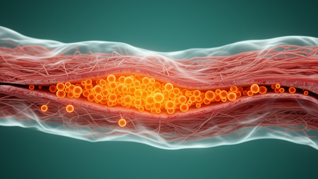
Image Credits: AI Generated
DOI: 10.1038/s44161-026-00881-9
Keywords: cardiac regeneration, succinate dehydrogenase, malonate, myocardial infarction, cardiomyocyte proliferation, fibrosis, epigenetics, H3K4me3, H3K27me3, metabolism, single-nucleus sequencing, myofibroblasts
Cite Scienmag News
Ophelia Keating. (October 8, 2026). Metabolic Switch in Heart Cells Rewinds Injury Into Regeneration, Study Finds. Scienmag. https://scienmag.com/metabolic-switch-in-heart-cells-rewinds-injury-into-regeneration-study-finds/
Ophelia Keating. "Metabolic Switch in Heart Cells Rewinds Injury Into Regeneration, Study Finds." Scienmag, 8 October 2026, https://scienmag.com/metabolic-switch-in-heart-cells-rewinds-injury-into-regeneration-study-finds/. Accessed 8 October 2026.
Ophelia Keating. "Metabolic Switch in Heart Cells Rewinds Injury Into Regeneration, Study Finds." Scienmag. October 8, 2026. https://scienmag.com/metabolic-switch-in-heart-cells-rewinds-injury-into-regeneration-study-finds/


