


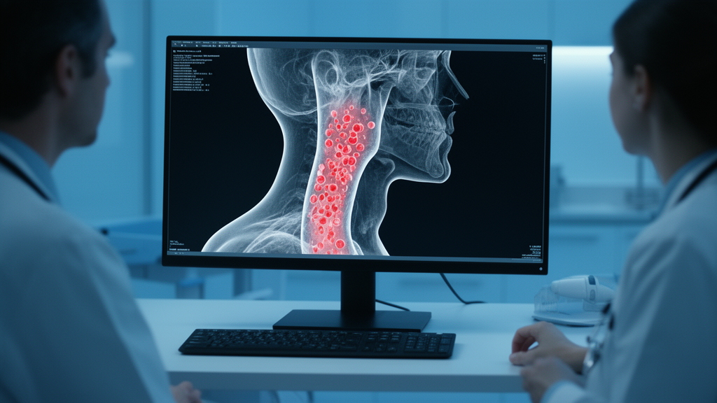
Narrowed arteries in the neck are responsible for roughly one in five ischemic strokes, but the plaques that actually rupture and trigger these events are notoriously hard to spot without specialized imaging. Now a pilot study from the University of Bern suggests that a combination of routine computed tomography angiography, ultrasound measurements, and computer simulations of blood flow could help identify these dangerous lesions in patients who lack access to the current gold standard, plaque magnetic resonance imaging.
The research, published in PLOS Digital Health by a team led by Shaokai Zheng and David Bervini, focused on so-called vulnerable plaques in carotid atherosclerosis. These are lesions prone to rupture that contain features such as intra-plaque hemorrhage, a lipid-rich necrotic core, or a ruptured fibrous cap. Once a plaque ruptures, thrombo-embolic debris can travel to the brain and cause a stroke. Detecting such plaques matters because their prevalence among patients with asymptomatic carotid stenosis is estimated at around 26 percent, and identifying them guides decisions about carotid endarterectomy, the surgical removal of plaque from the artery.
Today, the preferred way to assess plaque vulnerability is multi-contrast plaque MRI, which can visualize hemorrhage and cap integrity directly. But MRI scanners with the right sequences and the radiological expertise to interpret them are concentrated in tertiary centers, creating bottlenecks and diagnostic delays. The Bern team asked a different question: could the physics of blood flow itself, simulated on ordinary clinical data, reveal the fingerprint of a dangerous plaque?
Their workflow began with nine patients scheduled for carotid endarterectomy between January 2021 and March 2023, yielding eighteen carotid arteries for analysis, nine of which were rated vulnerable on plaque MRI according to the Plaque-RADS grading system. From each patient’s CT angiogram, the researchers segmented the carotid bifurcation, the fork where the common carotid artery splits into the internal and external branches, and reconstructed three-dimensional geometries containing between 1.3 and 1.8 million computational cells. Crucially, instead of imposing generic pulsatile waveforms, they anchored the simulations in reality by using peak systolic and diastolic flow velocities measured with neurovascular ultrasound, a standard first-line examination, to set the boundary conditions of the model.
The team then solved the Navier-Stokes equations for blood flow, modeled as a Newtonian fluid under laminar conditions, at both peak systole and diastole using steady-state simulations in COMSOL Multiphysics. Each simulation took under an hour on an ordinary desktop computer. From the resulting velocity fields, they extracted an unusually rich set of 289 features, including geometric measures such as bifurcation angles and stenosis ratios, and hemodynamic quantities such as wall shear stress, vorticity, and helicity, a measure of the corkscrew rotation of flow. Importantly, they went beyond simple averages, computing higher-order statistics such as skewness and kurtosis, which describe the asymmetry and tailedness of how these quantities are distributed inside the vessel.
Two machine learning classifiers, logistic regression and a linear support vector machine, were trained to distinguish vulnerable from non-vulnerable arteries. To guard against overfitting in this tiny dataset, the researchers used leave-one-out cross-validation with recursive feature elimination embedded inside each fold, preventing any data leakage, alongside correlation filtering and L2 regularization. The classifiers achieved mean areas under the receiver operating characteristic curve of 0.77 and 0.78 respectively, moderate discriminative performance with wide confidence intervals that reflect the small sample size.
The most striking result was which features the algorithms kept selecting. Mean helicity emerged as the strongest protective signal, with an odds ratio of 0.5, meaning more organized helical flow was associated with non-vulnerable plaques. Higher kurtosis of helicity, indicating concentrated and coherent helical flow structures, was likewise protective, while skewness of streamwise vorticity, which captures how swirling structures aligned with the vessel axis are distributed, ranked among the strongest risk predictors. Eight features were consistently chosen by both independent classifiers, lending credibility to the signal.
Equally telling was what did not matter. Wall shear stress, long the centerpiece of hemodynamic risk research, showed no significant differences between groups in univariate comparisons, and only one shear-related feature, the skewness of relative wall shear stress during diastole, was selected by a classifier. The authors argue this makes physical sense: wall shear stress quantification is exquisitely sensitive to imaging resolution, surface reconstruction choices, and near-wall velocity treatment, with reported deviations of up to 38 percent in severely stenotic cases. Bulk intravascular quantities like helicity, by contrast, are more robust to these uncertainties, a lesson already emerging from coronary flow studies.
Geometric features fared no better. Bifurcation angle, tortuosity, and diameter ratios, all previously proposed risk markers, showed no significant group differences and were largely ignored by the classifiers. The team notes that bifurcation angle measurements shift dramatically depending on where along the curving branches they are taken, underscoring the field’s need for standardized geometric definitions, or perhaps a shift away from geometry altogether toward flow-based biomarkers.
The clinical vision is a triage system rather than an MRI replacement. Because neurovascular ultrasound and CT angiography are already part of routine workups, and the simulation and analysis pipeline is largely automated, the workflow could pre-screen patients locally, referring only those flagged as high-risk for specialized plaque MRI while sparing low-risk patients unnecessary trips to tertiary centers. Sub-analyses excluding non-stenotic arteries preserved classifier performance, and the authors emphasize that the ability to correctly classify healthy contralateral vessels is practically important since both sides are routinely assessed.
Caution is warranted. This was a retrospective, single-center proof of concept with only eighteen arteries, all from surgical candidates, and the authors themselves frame the conclusions as hypothesis-generating rather than confirmatory. Prospective, multicentric validation with larger cohorts, automated segmentation, and eventually hard outcomes such as stroke incidence will be essential. Still, the idea that the statistics of swirling blood, computed from scans most clinics already possess, could whisper which plaques are about to turn lethal is a compelling glimpse of accessible, physics-driven stroke prevention.
Subject of Research: Using computational fluid dynamics from CT angiography and ultrasound with machine learning to identify vulnerable carotid plaques
Article Title: Data driven approach to identify vulnerable plaques in carotid atherosclerosis using computed tomography imaging and computational fluid dynamics: A pilot study
Article References: Zheng, S., Bervini, D., Besson, H., Martínez, S. G., Heldner, M. R., Müller, M., Meinel, T., Kaesmacher, J., Harloff, A., & Obrist, D. (2026). Data driven approach to identify vulnerable plaques in carotid atherosclerosis using computed tomography imaging and computational fluid dynamics: A pilot study. PLOS Digital Health, 5(10), e0001762. https://doi.org/10.1371/journal.pdig.0001762
Image Credits: AI Generated
DOI: 10.1371/journal.pdig.0001762
Keywords: carotid atherosclerosis, vulnerable plaque, computational fluid dynamics, CT angiography, neurovascular ultrasound, machine learning, stroke, plaque MRI, helicity, wall shear stress, hemodynamics, PLOS Digital Health
Cite Scienmag News
Audrey Campbell. (October 8, 2026). Blood Flow Simulations From Routine Scans Could Flag Dangerous Carotid Plaques Without MRI. Scienmag. https://scienmag.com/blood-flow-simulations-from-routine-scans-could-flag-dangerous-carotid-plaques-without-mri/
Audrey Campbell. "Blood Flow Simulations From Routine Scans Could Flag Dangerous Carotid Plaques Without MRI." Scienmag, 8 October 2026, https://scienmag.com/blood-flow-simulations-from-routine-scans-could-flag-dangerous-carotid-plaques-without-mri/. Accessed 8 October 2026.
Audrey Campbell. "Blood Flow Simulations From Routine Scans Could Flag Dangerous Carotid Plaques Without MRI." Scienmag. October 8, 2026. https://scienmag.com/blood-flow-simulations-from-routine-scans-could-flag-dangerous-carotid-plaques-without-mri/


