


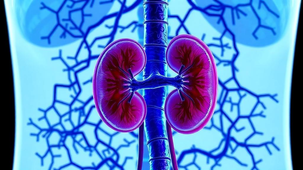
Diabetic nephropathy, the slow and relentless destruction of the kidneys that follows years of elevated blood sugar, remains the single leading cause of end-stage renal disease worldwide. Despite decades of research, the therapeutic arsenal against it remains thin: patients can slow the decline with tight glucose control, blood pressure management, and newer agents such as SGLT2 inhibitors and finerenone, but few treatments directly attack the molecular machinery that converts a functioning filter into scar tissue. Now a team of researchers at the First Affiliated Hospital of Harbin Medical University in Harbin, China, has identified a surprising culprit in that machinery: an RNA-binding protein called PTBP3, better known for its roles in cancer biology, which appears to orchestrate a critical injury program in the kidney’s most delicate cells. The study, published in Molecular Biology Reports, suggests that silencing PTBP3 can meaningfully protect kidney structure and function in diabetic mice.
The kidney’s filtering units, the glomeruli, depend on a specialized layer of cells called podocytes. These cells wrap around the glomerular capillaries with interdigitating foot processes, forming slit diaphragms that act as the final, exquisitely selective barrier preventing albumin and other precious proteins from leaking into the urine. Podocytes are terminally differentiated epithelial cells; once damaged, they do not readily regenerate. One of the most consequential forms of podocyte injury is a process known as epithelial-to-mesenchymal transition, or EMT, in which these highly specialized epithelial cells lose their characteristic architecture and adhesion molecules and begin expressing mesenchymal markers such as N-cadherin, vimentin, and fibronectin. The transformed cells become motile, detach from the glomerular basement membrane, and contribute to the fibrotic scarring that progressively chokes off renal function. Prior work has implicated the TGF-beta signaling pathway, and its intracellular mediator SMAD3, as a central driver of this transition in the diabetic kidney.
The Harbin team began not in the laboratory but in the data. By integrating transcriptomic datasets from public repositories, they searched for RNA-binding proteins that were aberrantly expressed in diabetic kidney tissue, and PTBP3 emerged as a standout candidate. Polypyrimidine tract-binding protein 3 is a member of a family of proteins that bind specific sequence motifs in messenger RNA molecules, influencing their stability, translation, and alternative splicing. PTBP3 has attracted increasing attention in oncology research, where it has been shown to promote epithelial-to-mesenchymal transition and metastasis in lung adenocarcinoma, drive growth in colorectal and gastric cancers, and modulate immune escape in gallbladder cancer. Its role in kidney disease, however, had remained essentially unexplored, making the new findings a notable expansion of the protein’s known biological territory.
To confirm that the computational signal reflected real biology, the researchers turned to animal and cell models. In mice made diabetic by injection of streptozotocin, a compound that selectively destroys insulin-producing beta cells and reliably induces hyperglycemia, PTBP3 levels rose in kidney tissue. The same elevation appeared in podocytes grown in culture and exposed to high glucose concentrations, the standard in vitro model of diabetic cellular stress. These convergent observations, in vivo and in vitro, established that PTBP3 upregulation is a reproducible feature of the diabetic kidney environment rather than an artifact of any single experimental system, and positioned the protein as a plausible participant in disease pathogenesis.
The decisive experiment came next. The team knocked down PTBP3 in diabetic mice and monitored the classic functional readouts of kidney injury. The results were striking: 24-hour urinary protein excretion, a direct measure of the damaged filtration barrier, was significantly reduced in the knockdown animals. Serum creatinine and blood urea nitrogen, the two workhorse clinical markers of declining kidney clearance, were also substantially attenuated compared with diabetic controls. In other words, removing this single RNA-binding protein preserved measurable kidney function in the face of ongoing diabetes.
Histological analysis told a matching structural story. Under the microscope, the kidneys of diabetic mice typically show glomerular hypertrophy, expansion of the mesangial matrix that crowds the filtering tuft, and the deposition of collagen-rich fibrotic tissue. In the PTBP3 knockdown animals, all of these pathological features were ameliorated. Renal fibrosis was reduced, and the glomerular architecture was better preserved. The researchers further demonstrated that PTBP3 silencing inhibited the epithelial-to-mesenchymal transition of podocytes in the diabetic mice, an effect visible at the molecular level as restored expression of E-cadherin, the epithelial adhesion molecule that anchors podocytes in place, alongside reduced expression of the mesenchymal markers N-cadherin, vimentin, and fibronectin. Cultured podocytes subjected to high glucose showed the same pattern, reinforcing the consistency of the mechanism across model systems.
The mechanistic core of the paper lies in how PTBP3 exerts these effects, and here the authors provide a satisfying molecular explanation centered on SMAD3, the transcriptional mediator of TGF-beta signaling that has long been associated with fibrotic disease. Using dual-luciferase reporter assays and RNA immunoprecipitation, the team showed that PTBP3 physically interacts with the 3-prime untranslated region of SMAD3 messenger RNA. This interaction stabilizes the transcript, increasing the total amount of SMAD3 protein produced and, consequently, the levels of its phosphorylated, active form. In effect, PTBP3 acts as a post-transcriptional amplifier: by protecting SMAD3 mRNA from degradation, it boosts the signaling output of the TGF-beta pathway, which in turn drives podocytes toward the mesenchymal, fibrosis-promoting state. When PTBP3 is removed, SMAD3 expression falls, phosphorylation declines, and the EMT program loses its principal engine.
This finding places PTBP3 within a growing map of post-transcriptional regulation in diabetic kidney disease. The SMAD3 axis itself is well established: genetic deletion of Smad3 has previously been shown to prevent renal fibrosis and inflammation in models of type 2 diabetic nephropathy, and multiple microRNAs and circular RNAs have been described that modulate SMAD3 expression in mesangial cells and podocytes. What the new study adds is a specific RNA-binding protein that tunes the pathway at the level of mRNA stability, a layer of control that has been harder to drug than receptors or kinases but is increasingly recognized as a rich source of therapeutic targets. The parallel with cancer biology is also instructive, since PTBP3 has been repeatedly linked to TGF-beta-driven EMT in tumors, suggesting that the protein operates a conserved program of epithelial plasticity that the diabetic kidney co-opts for pathological remodeling.
The clinical implications, while still distant, are worth considering carefully. Diabetic nephropathy affects an enormous fraction of the growing global population with diabetes, and a substantial share of patients progress to dialysis or transplantation despite current best care. A target that acts upstream of SMAD3, and that demonstrably reduces proteinuria and preserves histology in a diabetic mouse model, represents the kind of mechanistic entry point that could eventually yield new drug classes, whether antisense oligonucleotides, small molecules that disrupt the PTBP3-SMAD3 mRNA interaction, or delivery strategies aimed specifically at podocytes. The authors note that PTBP3 knockdown ameliorated renal fibrosis and preserved renal function, underscoring the therapeutic potential of targeting podocyte epithelial-mesenchymal transition in diabetic nephropathy. At the same time, the usual caveats apply: mouse models of streptozotocin-induced diabetes capture only part of human diabetic kidney disease, and PTBP3’s roles in other tissues, including its documented effects in hematopoietic and endothelial biology, will need to be weighed against any systemic intervention.
For now, the study stands as a clear demonstration that the fate of the kidney’s filtration barrier can be steered by a single post-transcriptional regulator. By tracing a line from public transcriptomic data through diabetic mice and glucose-stressed podocytes down to a specific interaction between an RNA-binding protein and the untranslated region of a fibrotic transcription factor, the Harbin researchers have connected a molecular mechanism to clinically meaningful outcomes: less protein in the urine, lower creatinine, less scarring, better-preserved glomeruli. If those findings can be replicated in additional models and extended toward human tissue validation, PTBP3 may join the short list of molecules that clinicians watch as potential handles on one of diabetes’ most feared complications. The work was supported by the Basic Scientific Research Business Expenses Project of Colleges and Universities in Heilongjiang Province, and all animal experiments were approved by the Ethics Committee of the First Affiliated Hospital of Harbin Medical University.
Subject of Research: The role of the RNA-binding protein PTBP3 in Smad3-dependent podocyte injury and fibrosis in diabetic nephropathy
Article Title: PTBP3 promotes diabetic nephropathy via Smad3-dependent podocyte-mesenchymal transition
Article References: Li, Y., Fu, Y., Wang, X., Ma, X., Cong, G., & Sui, M. (2026). PTBP3 promotes diabetic nephropathy via Smad3-dependent podocyte-mesenchymal transition. Molecular Biology Reports, 53(1), Article 1628. https://doi.org/10.1007/s11033-026-12788-6
Image Credits: AI Generated
DOI: 10.1007/s11033-026-12788-6
Keywords: diabetic nephropathy, PTBP3, podocytes, SMAD3, epithelial-mesenchymal transition, renal fibrosis, RNA-binding proteins, TGF-beta signaling, chronic kidney disease, proteinuria, mRNA stability, kidney disease
Cite Scienmag News
Drew Townsend. (October 7, 2026). RNA-Binding Protein PTBP3 Drives Kidney Scarring in Diabetes, Study Finds. Scienmag. https://scienmag.com/rna-binding-protein-ptbp3-drives-kidney-scarring-in-diabetes-study-finds/
Drew Townsend. "RNA-Binding Protein PTBP3 Drives Kidney Scarring in Diabetes, Study Finds." Scienmag, 7 October 2026, https://scienmag.com/rna-binding-protein-ptbp3-drives-kidney-scarring-in-diabetes-study-finds/. Accessed 7 October 2026.
Drew Townsend. "RNA-Binding Protein PTBP3 Drives Kidney Scarring in Diabetes, Study Finds." Scienmag. October 7, 2026. https://scienmag.com/rna-binding-protein-ptbp3-drives-kidney-scarring-in-diabetes-study-finds/


