


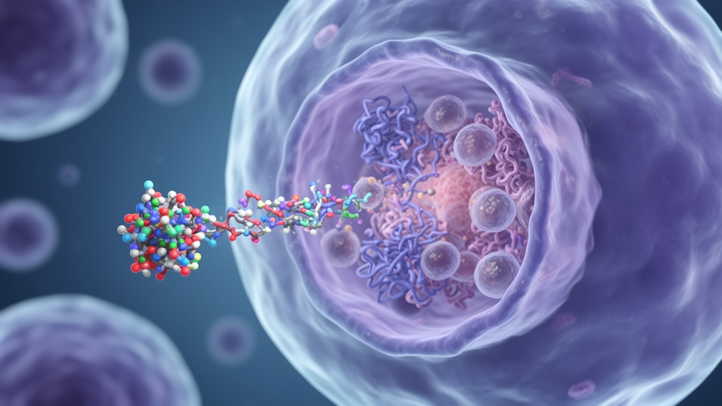
A 2026 review identifies HIV-1 viral protein R, or Vpr, as a compact multifunctional accessory protein that coordinates several processes associated with viral replication and disease progression. The 96-amino-acid protein can arrest infected cells at the G2/M phase, trigger mitochondrial apoptosis, alter host signaling, increase envelope expression and manipulate endogenous retroelements. Evidence assembled by Juan Du, Qingchen Li, Shao-Hua Wang, Yifei Zhao and Ke Zhao at First Hospital of Jilin University links these activities to the possible role of Vpr in AIDS pathogenesis and viral persistence, while emphasizing that most mechanistic findings come from cultured cells and have not been definitively shown to drive AIDS in people.
Vpr is one of four HIV-1-specific accessory proteins, alongside Vif, Nef and Vpu, that support viral replication and pathogenicity in vivo. First characterized in 1987, it is unusually conserved across HIV-1 strains and divergent primate lentiviruses. Loss-of-function mutants can regain full-length Vpr during prolonged transmission in primates or humans, indicating strong evolutionary selection for its function.
Structural studies, principally using nuclear magnetic resonance spectroscopy and later crystallography, have resolved three alpha-helical regions: residues 17–33, 40–48 and 55–83. The first two helices are largely polar, whereas helix 3 is amphipathic, allowing it to associate with lipid membranes and helping explain the perinuclear location of Vpr in host cells. The protein’s ability to manipulate the host ubiquitin-proteasome system is central to many of its effects.
Vpr acts as an adaptor for the CUL4-DDB1-DCAF1 ubiquitin ligase complex, commonly called CRLCul4. A crystal structure showed that helices 1 and 3 engage DCAF1, also known as Vpr-binding protein, while helix 2 contacts uracil-DNA glycosylase 2. This arrangement allows Vpr to recruit cellular proteins for proteasomal degradation.
Among the substrates are UNG2, exonuclease 1 and HLTF, all connected to DNA repair. Their depletion compromises genome maintenance, activates ATR- and Chk1/2-dependent checkpoint signaling and inhibits CDC25C, reducing cyclin B1-CDK1 activity and holding cells at G2/M. Vpr may also act directly on CDC25C and on regulators including WEE1, 14-3-3 proteins and PP2A through pathways that do not require the proteasome.
Cell-cycle arrest can benefit HIV-1 because the viral long terminal repeat, which contains the proviral promoter, is more active in G2-arrested cells. Vpr consequently boosts viral replication in proliferating cell lines and primary CD4+ T cells, whereas terminally differentiated macrophages already occupy G0 and do not show the same benefit. The same structural helix is central to another major function.
Vpr delivered by the incoming virion can insert partially into the outer mitochondrial membrane, dissipate membrane potential and release apoptogenic factors including apoptosis-inducing factor and cytochrome c. Cytochrome c then supports caspase-9 activation. Vpr also binds adenine nucleotide translocase, disturbing mitochondrial homeostasis and increasing reactive oxygen species, which can activate ATR-Chk1 DNA-damage signaling.
Experiments in Jurkat cells and peripheral blood mononuclear cells have linked this cytotoxicity to possible CD4+ T-cell depletion, but clinical validation remains limited. The coupling of G2 arrest and apoptosis creates a delicate evolutionary balance. Arrest can raise proviral transcription and favor production of progeny virions, whereas killing an infected producer cell may limit sustained viral output.
Studies cited in the review indicate that cell-cycle arrest generally precedes and peaks before strong apoptotic activity. Vpr may therefore calibrate the timing of transcriptional amplification and cellular death rather than simply promote one outcome. This distinction matters because the review concludes that no single established Vpr function has been validated as the driver of HIV-1 infection or AIDS.
Many findings derive from tumor-derived lines, primary cells, overexpression systems or laboratory infection models, and discrepancies often arise when findings from these systems are compared. The authors caution that physiological relevance cannot be assumed from an in vitro phenotype alone. Vpr can also enhance the expression and stability of the HIV-1 envelope protein, Env, particularly in macrophages, a major target cell and potential viral reservoir.
In primary macrophages, DCAF1 binding is required for Vpr-mediated Env upregulation. Vpr inhibits endosome-related degradation of Env by promoting CRLCul4-dependent proteasomal degradation of LAPTM5, a lysosomal transmembrane protein; loss of LAPTM5 reduces lysosomal clearance of Env and improves the infectivity and cell-to-cell transmission of newly formed virions among macrophages. Vpr also depletes the endonuclease Dicer and host factors including gelsolin and PU.1.
In macrophages, Vpr and Nef can reduce the surface mannose receptor, which otherwise destabilizes Env and impairs virion assembly and release. Together, these mechanisms may increase infectious virion production and facilitate transfer between macrophages and CD4+ T cells, although most detailed evidence remains from in vitro systems. These macrophage findings also connect Vpr to several routes of HIV-1 dissemination.
Phagocytosis of infected T cells can be more efficient than direct infection for delivering HIV-1 to macrophages, while infected macrophages can present Env-dependent virions on their surfaces and transmit virus to CD4+ T cells. Direct contact between infected T cells and uninfected macrophages can also promote membrane fusion and formation of multinucleated giant cells, which have been observed in infected patients and can be highly virus-productive. Vpr-mediated elevation of Env therefore has consequences beyond particle production: it may influence cell-to-cell spread, macrophage reservoirs and the efficiency of transfer between immune-cell compartments.
The review does not establish that these pathways are sufficient to sustain viral persistence in vivo, and it does not provide evidence for a clinical intervention targeting Vpr. Host-cell signaling offers another route by which Vpr may support viral replication. Virion-packaged Vpr activates nuclear factor of activated T cells without requiring the upstream calcium flux normally initiated by plasma-membrane receptors, allowing productive infection of Jurkat cells, primary T cells and macrophages.
Vpr also promotes proteasome-dependent degradation of interferon regulatory factor 3, a central component of type I interferon production. Its effects on nuclear factor kappa B are unresolved. Some studies report that Vpr and DCAF1 inhibit nuclear-kappa-B translocation, while others show activation through TAK1, IKKα/β, DNA-damage signaling or interactions with cell-cycle machinery.
Donor macrophages in one experiment differed markedly in their response, suggesting intrinsic biological variation. The review says the net effect remains to be validated even though the convergent cellular outcome may be enhanced proviral transcription and replication. Associations between particular Vpr variants and long-term nonprogressors, people who delay or avoid progression to AIDS, point to a potentially important role in pathogenesis.
Candidate variants include Q3R, N16H, T19A, Q65R, F72L/A, R77Q/A, R80A and R90N/K. Q65R and R80A disrupt the Vpr-DCAF1 interface and therefore impair recruitment of CRLCul4, potentially disabling numerous Vpr activities. Yet T19A and R90N retain wild-type activity across known functions, and R77Q, previously associated with weaker apoptosis, was later found in HUT78 cells to retain G2/M arrest and apoptosis.
Its apparent instability in a proviral expression system, together with stronger arrest and cytotoxicity at low expression, may indicate that some variants alter the timing of producer-cell death rather than simply removing a function. These mixed phenotypes reinforce the idea that Vpr’s pathogenic effects may arise from coordinated, context-dependent activities rather than one mechanism. Newer functions may help explain unresolved links between Vpr and disease progression.
Vpr can promote proteasomal depletion of the methylcytosine dioxygenase TET2, affecting epigenetic regulation of interleukin-6 expression; a clinical cohort has also associated high Vpr detection with increased interleukin-6 and aggravated AIDS pathogenesis. Vpr can deplete PU.1, which links Env expression to suppression of innate immune responses in macrophages, and target SLF2, a regulator of the SMC5-SMC6 complex, in CEM-CD4 and primary CD4+ T cells. Vpr also has context-dependent effects on LINE-1 retrotransposition.
HIV-1 infection increased LINE-1 activity in a Vif- and Vpr-dependent manner in Jurkat cells, and L1 DNA accumulated in infected primary CD4+ T cells. Extracellular Vpr enhanced L1 replication in mouse models and patient-derived samples, whereas intracellular Vpr directly bound LINE-1 ORF2p and inhibited its reverse-transcriptase activity. The review notes that evidence connecting LINE-1 modulation to AIDS progression is still lacking.
Overall, the synthesis presents Vpr as an evolutionarily conserved coordinator of replication, cytotoxicity, immune disruption and persistence, but not yet as a clinically proven causal target; questions remain about its activity in primary cells, donor-dependent signaling effects, the balance between viral amplification and infected-cell death, and whether any of its functions can be therapeutically separated from those needed to maintain viral fitness.
Subject of Research: Multifunctional roles of HIV-1 Vpr in viral replication, immune disruption and AIDS pathogenesis
Article Title: HIV-1 Vpr: a multifunctional accessory protein with potential contributions to AIDS pathogenesis and persistence
Article References: Du, J., Li, Q., Wang, S.-H., Zhao, Y., & Zhao, K. (2026). HIV-1 Vpr: a multifunctional accessory protein with potential contributions to AIDS pathogenesis and persistence. Frontiers in Microbiology, 17, Article 1971957. https://doi.org/10.3389/fmicb.2026.1971957
Image Credits: AI Generated
DOI: 10.3389/fmicb.2026.1971957
Keywords: HIV-1, Vpr protein, AIDS pathogenesis, viral replication, G2/M arrest, apoptosis, immune evasion, macrophages, LINE-1 retrotransposition, long-term nonprogressors, multifunctional, accessory
Cite Scienmag News
Kristina Jarvis. (October 20, 2026). Research report: HIV-1 Vpr: a multifunctional accessory protein with potential contributions to AIDS pathogenesis and persistence. Scienmag. https://scienmag.com/research-report-hiv-1-vpr-a-multifunctional-accessory-protein-with-potential-contributions-to-aids-pathogenesis-and-persistence/
Kristina Jarvis. "Research report: HIV-1 Vpr: a multifunctional accessory protein with potential contributions to AIDS pathogenesis and persistence." Scienmag, 20 October 2026, https://scienmag.com/research-report-hiv-1-vpr-a-multifunctional-accessory-protein-with-potential-contributions-to-aids-pathogenesis-and-persistence/. Accessed 20 October 2026.
Kristina Jarvis. "Research report: HIV-1 Vpr: a multifunctional accessory protein with potential contributions to AIDS pathogenesis and persistence." Scienmag. October 20, 2026. https://scienmag.com/research-report-hiv-1-vpr-a-multifunctional-accessory-protein-with-potential-contributions-to-aids-pathogenesis-and-persistence/


