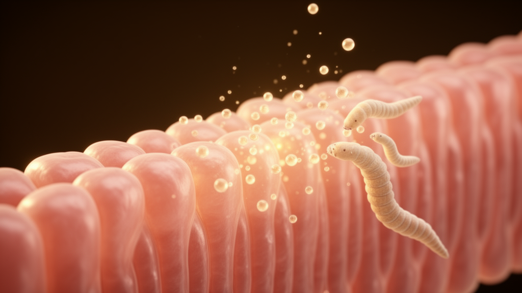
Human strongyloidiasis is a chronic, long-lasting infection caused by the helminth parasite Strongyloides stercoralis, a nematode notable for a peculiar autoinfective cycle that allows it to persist in the host for years. According to the authors, the interplay between the parasite and the host should entail mechanisms of adaptation to tolerate the chronic presence of this nematode. According to the authors of a new study, however, the mechanisms underpinning this interaction remain poorly understood, and their work was designed to add molecular detail to a picture that is still largely incomplete.
The team took a multi-omics approach, combining transcriptomic and proteomic readouts with measurements of secreted inflammatory signalling molecules, to examine how human intestinal epithelial cells respond to two distinct parasite-derived stimuli: live infective larvae, known as iL3, and extracellular vesicles, or EVs, released by those larvae. The iL3 used in the experiments were isolated from a clinical faecal sample and then maintained in vitro for up to one week, a period long enough for the parasites to release EVs into the surrounding conditioned medium. The researchers emphasise that all of this work was carried out in cell culture, using the Caco-2 human intestinal epithelial cell line, so the findings describe events in a controlled laboratory setting rather than in infected patients.
Caco-2 cells were exposed to either live larvae or their EVs for three different durations: 6 hours, 24 hours and 72 hours. The timing turned out to matter considerably. After just 6 hours of co-incubation, live iL3 induced the release of pro-inflammatory cytokines, specifically IL-8 and CXCL10. The EVs, by contrast, triggered the release of CXCL10 only, and at a lower intensity than that seen with live larvae. This early divergence hinted that the two stimuli, although derived from the same parasite stage, do not simply deliver the same signal in different doses.
The transcriptomic and proteomic analysis sharpened that distinction. Cells exposed to live larvae showed a pattern of cellular activation and a pro-inflammatory-like profile that built up over time and peaked 72 hours after exposure. The EVs behaved almost like a mirror image: their effects appeared earlier, with modulation of mRNA and protein expression occurring mostly at the 24-hour time point, and the direction of change was predominantly inhibitory, dampening cell activation, metabolism and immune responses. In other words, the soluble cargo of the vesicles appeared to push the epithelial cells away from activation while the living parasites pushed them toward it.
The authors interpret this asymmetry cautiously but suggestively. Because the EVs primarily down-modulated cell activation, they hypothesise that the vesicles may play a role in immune evasion, helping the parasite temper the very inflammatory pathways that live larvae provoke. They are careful to frame this as a hypothesis rather than a demonstrated mechanism, noting that more in-depth investigations are needed to validate the findings and to clarify what role parasite-derived EVs actually play in the pathophysiology of human strongyloidiasis.
Several limitations follow directly from the study design. The experiments used a single immortalised cell line rather than primary human intestinal tissue, a single parasite isolate obtained from one clinical sample, and a small set of time points. Dose relationships, the contribution of other parasite secretions present in the larval cultures, and the behaviour of the full immune response, which involves many cell types beyond the epithelium, all lie outside what this in vitro system can show. The authors themselves state that the current knowledge of the host-parasite interaction in strongyloidiasis remains limited and that their results are intended as novel insights rather than definitive answers.
Even with those caveats, the study offers a concrete molecular contrast that could guide future work. The authors state that more in-depth investigations are needed to validate these findings and to further elucidate the role of parasite-derived EVs in the pathophysiology of human strongyloidiasis. For now, the reported finding stands as an early, cell-culture-level observation: the same parasite presents its host’s intestinal cells with two very different molecular conversations, one loud and inflammatory, the other quieter and dampening, and the balance between them may help explain how this worm sustains its chronic residence.
Subject of Research: How Strongyloides stercoralis infective larvae and their extracellular vesicles modulate human intestinal epithelial cells in vitro
Article Title: In vitro investigation of the host-pathogen interaction in human strongyloidiasis at the intestinal level
Article References: Deiana, M., Padovani, N., Vezzelli, E., Manfredi, M., Locatelli, E., Moron Dalla Tor, L., Ronzoni, N., Ursini, T., Piubelli, C., Buonfrate, D., & Tiberti, N. (2026). In vitro investigation of the host-pathogen interaction in human strongyloidiasis at the intestinal level. Frontiers in Cellular and Infection Microbiology, 16, Article 1981291. https://doi.org/10.3389/fcimb.2026.1981291
Image Credits: AI Generated
DOI: 10.3389/fcimb.2026.1981291
Keywords: Strongyloides stercoralis, strongyloidiasis, extracellular vesicles, infective larvae, Caco-2 cells, transcriptomics, proteomics, CXCL10, IL-8, host-pathogen interaction, immune evasion, in vitro study
Cite Scienmag News
Kristina Jarvis. (October 28, 2026). Parasite Larvae and Their Vesicles Push Gut Cells in Opposite Directions in Lab Study. Scienmag. https://scienmag.com/parasite-larvae-and-their-vesicles-push-gut-cells-in-opposite-directions-in-lab-study/
Kristina Jarvis. "Parasite Larvae and Their Vesicles Push Gut Cells in Opposite Directions in Lab Study." Scienmag, 28 October 2026, https://scienmag.com/parasite-larvae-and-their-vesicles-push-gut-cells-in-opposite-directions-in-lab-study/. Accessed 28 October 2026.
Kristina Jarvis. "Parasite Larvae and Their Vesicles Push Gut Cells in Opposite Directions in Lab Study." Scienmag. October 28, 2026. https://scienmag.com/parasite-larvae-and-their-vesicles-push-gut-cells-in-opposite-directions-in-lab-study/