


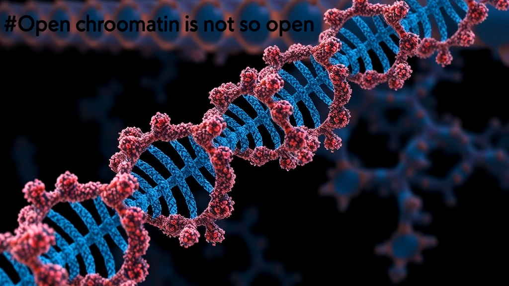
For decades, biology textbooks have drawn a deceptively simple picture of the genome’s physical state: dark, tightly packed heterochromatin silences genes, while light, airy euchromatin keeps active regions loose and accessible. A new study from researchers at the Massachusetts Institute of Technology, published in Nature Genetics, upends that tidy dichotomy. By combining high-resolution chromosome conformation data with polymer physics simulations, Joseph Paggi, Lawrence Long and Bin Zhang show that euchromatin is, in fact, largely condensed into compact domains whose densities rival those of heterochromatin. What distinguishes the active genome is not its overall looseness but the behavior of short regulatory stretches—promoters and enhancers—that protrude from the surfaces of these dense domains like handles on a suitcase, exposed and ready for the molecular machinery of transcription.
The technical hurdle the team faced is one that has haunted genome biologists for years: the three-dimensional arrangement of enhancers and promoters at the scale of individual nucleosomes—the fundamental spools around which DNA wraps—has remained essentially invisible. Popular chromosome conformation capture methods such as Hi-C average over millions of cells and blur out anything smaller than tens of kilobases. Super-resolution imaging can reach the nanoscale, but only for a handful of selected loci at a time. To bridge the gap, the researchers developed a simulation framework that leverages region-capture Micro-C contact maps, an assay that zooms in on megabase-scale windows with nucleosome-level resolution, to infer full conformational ensembles of genomic regions.
A central innovation of the work is a data-processing step the authors call neighbor balancing. Standard Micro-C analysis assumes that contact density is uniform across the genome, an assumption that quietly erases one of the most biologically meaningful signals: the fact that some stretches of DNA are simply more densely packed, and therefore more frequently in contact, than others. The new balancing strategy identifies variation in contact density instead of normalizing it away. When the team applied the method, sharp dips in contact density appeared precisely at active promoters and enhancers—places where the chromatin fiber opens up and nucleosomes thin out. These dips had been smoothed over in coarser, genome-wide datasets, but region-capture data preserved them, and neighbor balancing made them interpretable.
With the corrected contact maps in hand, the researchers turned to a maximum-entropy inversion approach, a technique rooted in statistical mechanics that finds the least-biased set of polymer structures consistent with the experimental constraints. The resulting simulated ensembles were then put through a battery of validation tests. The simulated structures reproduced pairwise distance distributions measured independently by chromatin tracing, a super-resolution imaging method that follows the positions of dozens of genomic loci in single cells. The simulations also contained packing domains and nucleosome clutches—discrete clusters of neighboring nucleosomes—matching structures that electron microscopy and super-resolution imaging studies had previously observed directly in cells. In other words, the model did not merely fit the data it was trained on; it recovered structural features seen by entirely different experimental techniques.
The most striking finding emerged when the team examined what those structures actually look like. Far from being uniformly open, euchromatin generally forms condensed domains with packing densities comparable to those of heterochromatin. The difference lies in scale: euchromatic domains are smaller than their heterochromatic counterparts, but the chromatin fiber inside them is nearly as tightly packed. Local nucleosome concentrations inside domain interiors frequently exceeded 400 micromolar, an extraordinarily crowded environment for DNA. This result aligns with a growing body of imaging evidence suggesting that so-called open chromatin is condensed but liquid-like in living cells, and it forces a rethinking of what the light and dark bands of classical cytology actually represent.
If euchromatin is mostly dense, how do genes get read? The answer, according to the simulations, lies in the geometry of the domain surfaces. Kilobase-scale regions at promoters and enhancers often protrude from the condensed domains, extending outward into the surrounding nuclear space where they become highly accessible. The team found that protrusion probability, the positions of clutch boundaries, and local contact density all correlate closely with ATAC-seq coverage, a genome-wide measure of chromatin accessibility. Where the fiber pokes out of a domain, transcription factors can find their binding sites; where it is buried inside, it cannot. Accessibility, in this picture, is not a bulk property of open chromatin but a surface phenomenon.
This arrangement effectively compartmentalizes regulatory elements from the surrounding chromatin, and the authors argue that the geometry serves a functional purpose. By lifting enhancers and promoters onto domain surfaces, the cell facilitates protein binding and enhancer–promoter communication, the long-range conversations between distant regulatory elements that switch genes on. The condensed cores may act as scaffolds that keep related regulatory elements in physical proximity, while the exposed protrusions provide the docking sites. The study also found that both packing domains and clutches tend to be smaller around ATAC-seq peaks, suggesting that local accessibility reshapes chromatin organization at multiple scales simultaneously.
The hierarchical picture that emerges runs from nucleosomes to clutches to domains, and it holds across species and cell types. The team validated their framework in mouse embryonic stem cells and in several human cell lines, finding condensed domains throughout. Even when the researchers examined data from cells depleted of cohesin, the ring-shaped protein complex that extrudes DNA loops, the local structure was largely maintained: chromatin expanded at large scales, but clutch-scale organization and the correlation between protrusions and accessibility persisted. This indicates that the condensed-domain architecture of euchromatin is not simply a byproduct of loop extrusion but reflects more fundamental physicochemical interactions within the chromatin fiber itself.
The implications reach well beyond structural biology. Misregulated enhancer–promoter communication underlies many developmental disorders and cancers, and drugs that target chromatin regulators, such as bromodomain inhibitors, are already in clinical use. A model in which accessibility depends on whether a regulatory element sits on a domain surface offers a concrete structural hypothesis for how such drugs work and why their effects are so context-dependent. It also reframes a long-standing question in the field: rather than asking how euchromatin stays open, biologists may now ask how specific regulatory elements are actively extruded or maintained on domain surfaces, and what molecular machinery performs that positioning.
The study’s computational framework, along with its simulation trajectories and analysis code, has been made publicly available, allowing other groups to apply the approach to their own region-capture datasets. As region-capture Micro-C spreads through the genomics community, the MIT team’s method could become a standard tool for converting contact maps into physically realistic, nucleosome-resolution structures. What began as a technical exercise in matrix balancing has delivered a conceptual shift: the active genome is not a loose tangle waiting to be read, but a dense, well-organized material whose most important working parts hang, deliberately and accessibly, on the outside.
Subject of Research: Three-dimensional organization of euchromatin and regulatory elements at nucleosome resolution
Article Title: Euchromatin forms condensed domains with short active regions on the surface
Article References: Paggi, J. M., Long, L. Y., & Zhang, B. (2026). Euchromatin forms condensed domains with short active regions on the surface. Nature Genetics. https://doi.org/10.1038/s41588-026-02775-9
Image Credits: AI Generated
DOI: 10.1038/s41588-026-02775-9
Keywords: chromatin, euchromatin, heterochromatin, nucleosomes, Micro-C, enhancers, promoters, gene regulation, 3D genome organization, polymer simulations, chromatin accessibility, super-resolution imaging
Cite Scienmag News
Juliet Wilcox. (September 25, 2026). Open Chromatin Is Not So Open: Active Genes Ride on Condensed Domains. Scienmag. https://scienmag.com/open-chromatin-is-not-so-open-active-genes-ride-on-condensed-domains/
Juliet Wilcox. "Open Chromatin Is Not So Open: Active Genes Ride on Condensed Domains." Scienmag, 25 September 2026, https://scienmag.com/open-chromatin-is-not-so-open-active-genes-ride-on-condensed-domains/. Accessed 4 October 2026.
Juliet Wilcox. "Open Chromatin Is Not So Open: Active Genes Ride on Condensed Domains." Scienmag. September 25, 2026. https://scienmag.com/open-chromatin-is-not-so-open-active-genes-ride-on-condensed-domains/


