


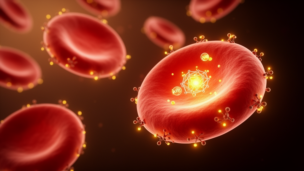
When the body runs low on oxygen, a molecular emergency switch flips on inside developing red blood cells. Scientists have now mapped a key part of that circuitry, showing how an energy-sensing enzyme called AMPK helps immature red cells mature under the stress of anemia by protecting a master regulator of oxygen metabolism. The findings, published in Frontiers in Physiology, trace a signaling chain that runs from the cell’s fuel gauge to the protein HIF-2α, and they point to new ways of thinking about anemias that fail to respond to standard drugs.
Anemia affects more than two billion people worldwide, making it one of the most common medical conditions on the planet. For many patients, treatment relies on erythropoiesis-stimulating agents, or ESAs, drugs that mimic the hormone erythropoietin and push the bone marrow to produce red blood cells. But a substantial fraction of patients, particularly those with chronic kidney disease or inflammatory disorders, develop ESA resistance, and their anemia persists despite escalating doses. Understanding the internal signals that govern red cell production under stress has therefore become a pressing goal for researchers seeking alternatives.
At the center of the new study is HIF-2α, a transcription factor that orchestrates the cellular response to low oxygen. HIF-2α drives the expression of genes involved in erythropoiesis, iron metabolism, and oxygen transport, and its stability is normally controlled by the von Hippel-Lindau protein, or VHL, which tags HIF-2α for destruction when oxygen is plentiful. When oxygen levels drop, VHL loses its grip, HIF-2α accumulates, and the cell activates its hypoxic program. The question the researchers asked was whether the cell’s energy status, sensed independently of oxygen, could also influence this process in red cell precursors.
To find out, the team used UT-7/EPO cells, a human cell line that depends on erythropoietin for survival and serves as a model for red cell progenitors. They cultured the cells under 1 percent oxygen with reduced erythropoietin, a combination designed to mimic the hostile environment that bone marrow faces during anemic stress. Under these conditions, cell viability dropped, apoptosis increased, and the cells ramped up expression of erythroid markers, reflecting an attempt to differentiate under pressure. The researchers then manipulated the AMPK and mTOR pathways with pharmacological agents to see how each affected the cells’ response.
AMPK, or AMP-activated protein kinase, acts as the cell’s fuel gauge. When energy supplies run low, AMPK switches on and shifts the cell into conservation mode, simultaneously inhibiting mTOR, a growth-promoting kinase that consumes substantial energy. The experiments showed that anemic stress activated AMPK and suppressed mTOR in the red cell precursors, and that this shift had a direct consequence for HIF-2α: the protein became markedly more stable, with a longer half-life than under normal conditions.
The mechanism behind that stabilization proved to be surprisingly direct. Using co-immunoprecipitation, the researchers found that when AMPK was activated with the drug AICAR, the physical interaction between HIF-2α and VHL weakened, effectively shielding HIF-2α from degradation. AICAR treatment also increased phosphorylation of HIF-2α at a specific site, serine 672, suggesting that this chemical modification may be the molecular handle by which AMPK disrupts the VHL binding. When the researchers instead forced mTOR into overdrive with the activator MHY1485, the protective effect disappeared, HIF-2α destabilized, and the cells lost their ability to mature properly.
Beyond stabilizing HIF-2α, AMPK activation triggered a broader program of erythroid differentiation and iron handling. The treated cells increased expression of the erythropoietin receptor, EPOR, along with DMT1 and FPN1, two proteins that respectively import iron into cells and export it after incorporation into heme. At the same time, levels of hepcidin, the hormone that restricts iron release from stores, and ferritin, the intracellular iron storage protein, both fell, freeing iron for hemoglobin synthesis. Intracellular iron levels recovered, supporting the cells’ push toward maturation.
Crucially, the researchers demonstrated that these downstream effects depended on HIF-2α itself. When they knocked down HIF-2α, the benefits of AMPK activation vanished: iron-related genes no longer responded, hepcidin and ferritin failed to drop, and erythroid differentiation stalled. Similarly, when mTOR was forced into overactivation, the entire cascade collapsed. The results establish AMPK, mTOR, and HIF-2α as a coherent signaling axis rather than three loosely connected players, with AMPK acting upstream and HIF-2α serving as the effector that translates energy stress into a red cell production program.
The clinical implications are notable. If the AMPK/mTOR/HIF-2α pathway controls stress erythropoiesis, drugs that modulate these kinases could offer a route to treating anemias that do not respond to erythropoietin-based therapies. Agents that activate AMPK, or that prevent mTOR from overriding the hypoxic response, might restore HIF-2α stability in patients whose red cell precursors have lost the ability to mature under stress. The iron-handling changes the researchers observed, particularly the reduction in hepcidin, could also complement existing strategies aimed at improving iron availability in chronic disease anemia.
As with any cell culture study, the findings will need validation in animal models and eventually in patients before they translate into treatments. The UT-7/EPO model, while useful, cannot fully reproduce the complexity of bone marrow physiology, and the interplay between AMPK signaling, systemic iron regulation, and inflammatory pathways in living organisms may introduce additional layers of control. Nevertheless, the study provides a clear mechanistic framework linking cellular energy status to red cell production, and it identifies specific molecular targets, including the HIF-2α-VHL interaction and serine 672 phosphorylation, that could guide drug development for one of medicine’s most persistent challenges.
Subject of Research: How AMPK activation stabilizes HIF-2α by disrupting VHL binding to promote erythroid differentiation and iron homeostasis under anemic stress via mTOR signaling
Article Title: AMPK activation stabilizes HIF-2α by disrupting VHL binding and promotes erythroid differentiation under anemic stress via the mTOR pathway
Article References: Cen, Y., Cai, P., Jiang, J., Huang, L., & Hua, Y. (2026). AMPK activation stabilizes HIF-2α by disrupting VHL binding and promotes erythroid differentiation under anemic stress via the mTOR pathway. Frontiers in Physiology, 17, Article 1932780. https://doi.org/10.3389/fphys.2026.1932780
Image Credits: AI Generated
DOI: 10.3389/fphys.2026.1932780
Keywords: AMPK, anemia, erythropoiesis, HIF-2α, mTOR, VHL, iron metabolism, hepcidin, erythroid differentiation, hypoxia, ESA resistance, red blood cells
Cite Scienmag News
Drew Townsend. (October 17, 2026). How a Cellular Fuel Sensor Helps Red Blood Cells Survive Anemia. Scienmag. https://scienmag.com/how-a-cellular-fuel-sensor-helps-red-blood-cells-survive-anemia/
Drew Townsend. "How a Cellular Fuel Sensor Helps Red Blood Cells Survive Anemia." Scienmag, 17 October 2026, https://scienmag.com/how-a-cellular-fuel-sensor-helps-red-blood-cells-survive-anemia/. Accessed 17 October 2026.
Drew Townsend. "How a Cellular Fuel Sensor Helps Red Blood Cells Survive Anemia." Scienmag. October 17, 2026. https://scienmag.com/how-a-cellular-fuel-sensor-helps-red-blood-cells-survive-anemia/


