


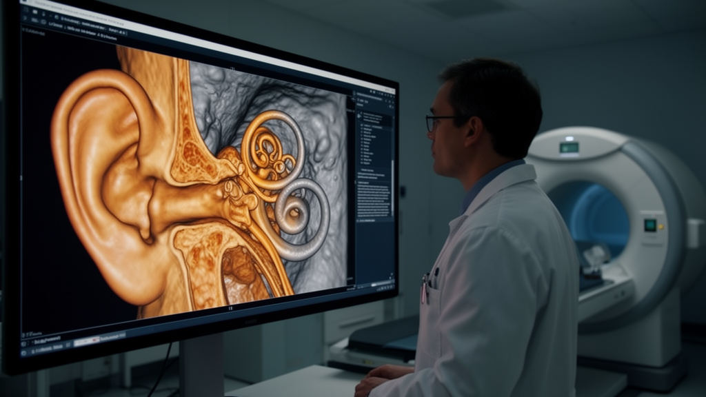
Deep inside the temporal bone—the densest bone in the human body—sits some of the most intricate anatomy in medicine. The cochlea, the facial nerve, three tiny ossicles, and a lattice of canals and windows all fit within a structure roughly the size of a walnut. Imaging this region well has always been a technical challenge, and now a large retrospective study from Jena University Hospital in Germany suggests that dynamic flat-panel computed tomography, better known as cone-beam CT, can do it with remarkable diagnostic performance while delivering a fraction of the radiation dose of conventional CT.
The research, published in Frontiers in Audiology and Otology, analyzed cone-beam CT scans of 216 patients covering 291 temporal bones, performed at the tertiary university center between 2015 and 2018. The cohort had a median age of 58 years and was 50.9 percent women. The team divided the scans into three groups: 58 patients with completely healthy temporal bones, 108 with middle or inner ear diseases, and 50 who had received a cochlear implant. A single radiology-trained observer graded the depiction quality of 24 distinct middle and inner ear structures on a three-point scale, ranging from not visible to clearly defined.
The technical setup matters. All scans were acquired on the same angiography unit, an Artis Q system from Siemens Healthineers, which repurposes the flat-panel detector of an interventional angiography suite for volumetric imaging. Instead of a rotating fan beam and multi-row detector array, cone-beam CT sweeps a cone-shaped X-ray beam around the head once, capturing projections on a large flat detector and reconstructing them into a three-dimensional volume. The median slice thickness across the cohorts was just 0.5 millimeters, and the median slice distance was 0.5 millimeters—although in the healthy cohort, where the ear was only an incidental imaging region, the median slice distance was 1 millimeter.
One of the study’s most counterintuitive findings was that diseased temporal bones were actually imaged better than healthy ones. Several middle and inner ear structures, including the geniculate ganglion, all three segments of the facial nerve, the chorda tympani, the tympanic membrane, the oval and round windows, the cochlear aqueduct, the helicotrema, the semicircular canals, the vestibule, and the vestibular aqueduct, were depicted with significantly higher quality in the pathology cohort than in healthy temporal bones, with p values below 0.01. Only the sigmoid sinus was identified more accurately in the healthy group.
The explanation is partly protocol and partly physics. The pathology cohort was scanned with a dedicated high-resolution protocol using significantly thinner slices, a mean of 0.48 versus 0.91 millimeters, and smaller image spacing, a mean of 0.50 versus 1.22 millimeters. In addition, pathology-related changes such as inflammatory opacification or soft-tissue filling in the middle ear can increase local contrast, delineating the borders of ossicles, the facial nerve canal, and the round window more clearly than in a completely air-filled healthy ear. Linear regression confirmed the protocol effect: in the healthy cohort, 17 of the 24 anatomical structures showed highly significant negative regression coefficients with slice thickness, meaning image quality rose as slices got thinner. The labyrinthine segment of the facial nerve, the oval window, and the round window had regression coefficients near −0.96 to −0.97. Image spacing showed the same pattern, with the semicircular canals at −0.91 and the helicotrema at −0.77.
In the diseased cohort, by contrast, slice thickness and spacing had little additional effect on most structures, likely because the scans were already performed at a uniformly fine 0.5-millimeter resolution, leaving little room for improvement. Notably, gender had no significant impact on depiction quality in either cohort, and across the four diagnostic subgroups—inflammatory, functional, malformation, and neoplastic—only the eardrum showed a significant difference between groups. That means essentially every structure, regardless of diagnosis, was equally usable for diagnostics.
The third cohort turned the technology toward quality control after cochlear implantation. Cone-beam CT enabled reliable assessment of the angular insertion depth of the electrode array, with a mean of 392 degrees, corresponding to slightly more than one full turn through the cochlear spiral, and a median of 399 degrees. The smallest measured insertion depth was 0 degrees, equivalent to incorrect positioning, and the largest was 732 degrees. The scans also allowed the team to determine the scalar location of the electrodes: in 78 percent of cases the electrode sat correctly in the scala tympani, in 10 percent it was misplaced in the scala vestibuli, in 8 percent it traversed both scalae, and in 4 percent it was not located in the cochlea at all—one instance involved electrode compression and the other a misimplantation into the eustachian tube.
There was one caveat on the implant side. While cone-beam CT excelled at measuring electrode position and insertion depth, the sensitivity and specificity values for determining the surgical access route—cochleostomy in 52 percent of cases versus the round window in 46 percent—were not strong enough to recommend the method for that specific purpose. Detection of cochleostomy had a sensitivity of 68.2 percent and a specificity of 60.7 percent, while round window detection yielded a sensitivity of 73.9 percent and a specificity of 44.4 percent. Newer cone-beam CT algorithms do, however, eliminate metallic artifacts more effectively than before, which is precisely why each individual electrode contact could be identified in the scans, a task where conventional CT often struggles.
The dose advantage is the headline number. Previous studies cited by the authors indicate that the effective dose at the petrous bone can be reduced by 40 to 45 percent compared with conventional CT while achieving better image quality, and ideally only 6 to 16 percent of the conventional CT dose is needed for consistently good images. The device theoretically supports slice thicknesses down to 0.14 millimeters, which would exceed the resolution of both conventional CT and temporal bone MRI, while saving time compared with MRI. Cone-beam CT is also quick to set up, mobile, and can be used for guidance during surgery itself, since patients under anesthesia produce negligible motion artifacts in the head region.
The authors acknowledge limitations: the evaluation was performed by a single observer, the retrospective design predetermined the diagnostic distribution, the healthy scans were not targeted at the ear as the primary region, and the malformation and neoplasia subgroups contained very few cases, limiting statistical power. Still, the overall conclusion is clear. Cone-beam CT demonstrates high diagnostic performance in detecting anatomical structures within the temporal bone, with better visualization in diseased ears than in healthy ones, establishing its suitability as a diagnostic method. It is also a reliable, low-dose tool for postoperative cochlear implant electrode position control—potentially allowing immediate quality control, and if necessary correction, directly in the operating room.
Subject of Research: Diagnostic performance of cone-beam CT for temporal bone anatomy and cochlear implant control
Article Title: High diagnostic performance of dynamic flat-panel computed tomography (Cone-Beam-CT) in detecting anatomical structures of temporal bones of healthy probands and patients with ear diseases
Article References: Podzimek, J., Hansch, A., Aschenbach, R., Teichgräber, U., Guntinas-Lichius, O., & Kouka, M. (2026). High diagnostic performance of dynamic flat-panel computed tomography (Cone-Beam-CT) in detecting anatomical structures of temporal bones of healthy probands and patients with ear diseases. Frontiers in Audiology and Otology, 4, Article 1857060. https://doi.org/10.3389/fauot.2026.1857060
Image Credits: AI Generated
DOI: 10.3389/fauot.2026.1857060
Keywords: cone-beam CT, temporal bone, cochlear implant, radiology, hearing loss, medical imaging, flat-panel CT, High, diagnostic, performance, dynamic, flat-panel
Cite Scienmag News
Ophelia Keating. (October 17, 2026). Cone-beam CT reveals ear anatomy in striking detail at a fraction of the radiation dose. Scienmag. https://scienmag.com/cone-beam-ct-reveals-ear-anatomy-in-striking-detail-at-a-fraction-of-the-radiation-dose/
Ophelia Keating. "Cone-beam CT reveals ear anatomy in striking detail at a fraction of the radiation dose." Scienmag, 17 October 2026, https://scienmag.com/cone-beam-ct-reveals-ear-anatomy-in-striking-detail-at-a-fraction-of-the-radiation-dose/. Accessed 17 October 2026.
Ophelia Keating. "Cone-beam CT reveals ear anatomy in striking detail at a fraction of the radiation dose." Scienmag. October 17, 2026. https://scienmag.com/cone-beam-ct-reveals-ear-anatomy-in-striking-detail-at-a-fraction-of-the-radiation-dose/


