A humble polyphenol found in tea leaves, grapes, oak bark, and countless other plants is drawing fresh attention from cancer researchers after a new preclinical study showed that orally administered gallic acid significantly shrank solid tumors in mice without the blood-damaging side effects that accompany standard chemotherapy. The research, published in the journal Medical Oncology, offers some of the most detailed in vivo evidence to date that this widely available plant compound can suppress tumor growth through clearly defined molecular mechanisms while leaving the immune system, liver, and kidneys essentially untouched.
The study, led by Greice Itamaro Heiden and colleagues at the Federal University of Santa Catarina in Brazil, set out to answer a question that has lingered over years of laboratory work: does gallic acid’s well-documented anticancer activity in cell cultures actually translate into a living organism, where tumors interact with blood vessels, immune cells, and a host of systemic factors that a petri dish cannot replicate? Most previous evidence came from in vitro experiments on epithelial and blood-derived cancer cell lines, including small-cell lung carcinoma, gastric adenocarcinoma, breast adenocarcinoma, promyelocytic leukemia, and glioma cells. Those studies showed that gallic acid can trigger cell death, halt the cell cycle, and interfere with signaling pathways that drive metastasis and angiogenesis. But the compound’s effects on mesenchymal solid tumors in a whole animal, and its safety profile compared head-to-head with a frontline chemotherapy drug, had never been systematically mapped.
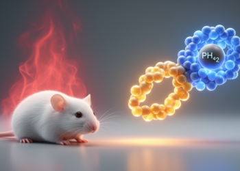
To fill that gap, the team turned to the Sarcoma 180 model, a transplantable mouse tumor that grows rapidly and is widely used for initial screening of potential anticancer agents. Fifty adult male Swiss mice received subcutaneous injections of S-180 ascitic cells, prepared at a standardized concentration of two million viable cells per animal after careful counting with Trypan Blue staining and a Neubauer chamber. Twenty-four hours later, the animals were randomly divided into five groups of ten. Three groups received purified gallic acid, at least 97.5 percent pure, by oral gavage once daily for seven consecutive days at doses of 10, 25, or 50 milligrams per kilogram of body weight. A fourth group received the chemotherapy drug 5-fluorouracil at 25 milligrams per kilogram intraperitoneally, while a control group received only the vehicle solution.
When the animals were euthanized on the eighth day and their tumors weighed, the results showed a clear dose-dependent effect. Mice treated with 25 milligrams per kilogram of gallic acid carried tumors weighing 9.38 grams per 100 grams of body weight, compared with 11.49 grams in the untreated control group, translating to a tumor growth inhibition of roughly 17.6 percent. At the highest dose of 50 milligrams per kilogram, tumor weight fell to 7.21 grams per 100 grams of body weight, an inhibition of 33.7 percent that reached high statistical significance. The lowest dose, 10 milligrams per kilogram, produced no significant effect. As expected, 5-fluorouracil remained the more powerful tumor killer, shrinking tumors by nearly 63 percent, but that potency came at a price the natural compound did not pay.
To understand how gallic acid was attacking the tumors, the researchers performed detailed histopathological and immunohistochemical analyses on the excised tissue. Under the microscope, the S-180 tumors displayed the aggressive features typical of this model: solid sheets of neoplastic cells infiltrating skeletal muscle and fat, perineural invasion, extensive coagulative necrosis, and high mitotic activity exceeding four mitoses per ten high-power fields. The tumor cells showed striking heterogeneity, with rhabdoid, epithelioid, spindle-shaped, and even unusual plant-like forms. Necrotic areas were visibly smaller in both the gallic acid and 5-fluorouracil groups, an early hint that both treatments were pushing tumor cells toward death.
The molecular story that emerged from the immunohistochemistry was remarkably coherent. Gallic acid at 50 milligrams per kilogram cut the expression of Ki-67, a nuclear protein that serves as a standard gauge of cell proliferation, from 14.2 percent of positive nuclei in controls to 9.5 percent. At the same time, it dramatically shifted the balance of the Bcl-2 protein family, which acts as the gatekeeper of mitochondrial suicide: the pro-apoptotic protein Bax rose sharply while the anti-apoptotic Bcl-2 fell, and levels of cleaved caspase-3, the executioner enzyme that dismantles the cell from within, increased significantly. Crucially, cleaved caspase-8, the initiator of the so-called extrinsic death receptor pathway, remained unchanged. Together, this pattern points to preferential activation of the intrinsic, or mitochondrial, apoptotic pathway, in which permeabilization of the mitochondrial membrane releases factors that trigger the caspase cascade and condemn the cell to programmed death.
The study also revealed a third strand in gallic acid’s mechanism: suppression of nuclear factor kappa B, or NF-κB, a transcription factor that functions as a master regulator of tumor cell survival, inflammation, and proliferation. In control animals, 38.4 percent of tumor cell nuclei stained positive for NF-κB; after gallic acid treatment, that figure dropped to 14.4 percent. Because NF-κB drives the production of anti-apoptotic proteins and promotes cell-cycle progression, its inhibition dovetails neatly with the observed increase in apoptosis and the decline in proliferation. The finding is consistent with earlier reports that gallic acid interferes with the MAPK/NF-κB pathway, possibly by reducing phosphorylation and acetylation of the p65 subunit, thereby dampening the factor’s transcriptional output. In effect, the compound appears to attack the tumor from multiple directions at once, strangling growth signals while simultaneously opening the doors to mitochondrial self-destruction.
The safety comparison with 5-fluorouracil may prove to be the study’s most consequential contribution. While the chemotherapy drug achieved greater tumor shrinkage, it devastated the animals’ white blood cell counts, dropping total leukocytes to 2.07 thousand per cubic millimeter, a hallmark of myelosuppression, and caused visible atrophy of the spleen’s white pulp, with shrunken follicles and disrupted lymphoid architecture. Gallic acid, by contrast, preserved leukocyte counts entirely. Liver and kidney weights were unchanged across all groups, serum levels of the liver enzymes AST and ALT stayed within normal ranges, and markers of renal function, urea and creatinine, showed no abnormalities. Histological examination confirmed normal liver and kidney architecture in the gallic acid group. The only notable biochemical shift, a lower AST level in the treated animals, likely reflects reduced tumor burden and less associated muscle damage rather than organ toxicity.
The authors are careful to frame these results as preclinical evidence rather than a promise of a new drug. The S-180 model, though fast and convenient, does not capture the full complexity of human malignancies, and the seven-day treatment window represents only an early snapshot of therapeutic potential. The antitumor efficacy of gallic acid, at roughly half that of 5-fluorouracil, would need substantial improvement, perhaps through combination regimens, novel formulations, or optimized dosing, before human trials could be contemplated. Nevertheless, the mechanistic clarity of the findings is striking: a single dietary polyphenol, taken orally, simultaneously suppressed proliferation, silenced a pro-survival transcription factor, and preferentially ignited the mitochondrial apoptotic pathway, all without measurable harm to blood, liver, or kidney. That combination of efficacy and safety is precisely what makes adjuvant candidates attractive, since the ideal partner to conventional chemotherapy would be one that adds tumor-killing power while shielding patients from the immunosuppression that limits standard treatment. As research into plant-derived anticancer compounds accelerates worldwide, gallic acid has now moved from the cell culture dish into the ranks of molecules with credible, mechanism-backed in vivo evidence, and the next chapter of its story will depend on whether that mitochondrial trigger can be pulled hard enough, and safely enough, in the clinic.
Subject of Research: Antitumor activity and safety of orally administered gallic acid in the murine Sarcoma 180 cancer model
Article Title: Antitumor efficacy and safety profile of gallic acid in the murine sarcoma 180 model
Article References: Heiden, G. I., Costa, M. A. D. S., Alfaia Silva, C., Carvalho, F. M. D. A. D., Amaral, R. G., Rivero, E. R. C., Modolo, F., Gondak, R., & de Albuquerque-Júnior, R. L. C. (2026). Antitumor efficacy and safety profile of gallic acid in the murine sarcoma 180 model. Medical Oncology, 43(11), Article 331. https://doi.org/10.1007/s12032-026-03455-8
Image Credits: AI Generated
DOI: 10.1007/s12032-026-03455-8
Keywords: gallic acid, Sarcoma 180, apoptosis, NF-κB, Ki-67, 5-fluorouracil, polyphenols, preclinical study, mitochondrial pathway, myelosuppression, cancer therapy, natural compounds
Cite Scienmag News
Nathaniel Bowman. (October 10, 2026). Gallic Acid Shrinks Tumors in Mice While Sparing Blood Cells, Study Finds. Scienmag. https://scienmag.com/gallic-acid-shrinks-tumors-in-mice-while-sparing-blood-cells-study-finds/
Nathaniel Bowman. "Gallic Acid Shrinks Tumors in Mice While Sparing Blood Cells, Study Finds." Scienmag, 10 October 2026, https://scienmag.com/gallic-acid-shrinks-tumors-in-mice-while-sparing-blood-cells-study-finds/. Accessed 10 October 2026.
Nathaniel Bowman. "Gallic Acid Shrinks Tumors in Mice While Sparing Blood Cells, Study Finds." Scienmag. October 10, 2026. https://scienmag.com/gallic-acid-shrinks-tumors-in-mice-while-sparing-blood-cells-study-finds/