


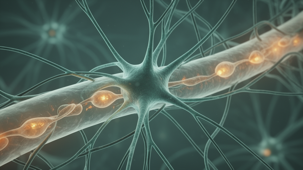
Researchers at Dali University have identified a distinctive astrocyte state characterized by high expression of extracellular matrix (ECM) and perineuronal net (PNN) signatures in major depressive disorder (MDD), along with three genes that show consistent disease-linked expression changes in both brain tissue and peripheral blood. The study, published as an original research article in Frontiers in Molecular Neuroscience, integrates bulk and single-cell transcriptomic datasets to map how ECM- and PNN-associated cellular states may contribute to the impaired neural plasticity long associated with depression. ECM and PNNs are structural components of the brain's extracellular environment that stabilize synapses and maintain extracellular homeostasis.
Although these structures are known regulators of neural circuit stability, the specific cellular states and molecular signatures tied to ECM and PNN alterations in depression had remained poorly understood, motivating the integrative approach taken by the team led by Yifan Wang, Chenrui Liang, and their colleagues. The researchers began by scoring astrocyte populations across multiple ECM and PNN signature metrics. Among the astrocyte clusters they examined, one cluster—designated Cluster2—stood out for exhibiting overall high ECM and PNN signature scores.
To move from cellular states to candidate molecules, the team integrated co-expression networks associated with Cluster2 with lists of differentially expressed genes linked to MDD. This cross-referencing yielded 28 overlapping genes, from which three were prioritized as cross-tissue candidate signatures: ACSL6, FIP1L1, and ZNF709. Critically, these three genes showed consistent expression trends across both brain and peripheral blood datasets, a property that makes them potentially useful as biomarkers accessible beyond brain tissue itself.
The direction of expression change differed among the three genes. ACSL6 and ZNF709 were downregulated in MDD, whereas FIP1L1 was upregulated. Functional enrichment analyses implicated all three genes in metabolic and amino-acid-related processes, while FIP1L1 showed additional associations with cell adhesion, cytoskeletal organization, and synaptic signaling.
On the strength of these functional links, the authors highlight FIP1L1 as a functionally prioritized candidate connected to adhesion- and synaptic-plasticity-related programs. Beyond gene-level findings, the study employed cell–cell communication analysis, pseudotime trajectory modeling, and in silico perturbation experiments to place the astrocyte findings in biological context. The communication analyses suggested altered intercellular signaling in the MDD-associated cellular environment, while the pseudotime analyses indicated increasing ECM and PNN activity across inferred astrocyte state transitions.
Together, these results support an astrocyte-centered working model in which a shift toward an ECM/PNN-high state accompanies the disease. To test whether the identified genes respond to stress at the cellular level, the team used a glucocorticoid-induced neuronal stress paradigm: HT22 cells treated with corticosterone (CORT). In this experimental system, ACSL6 and ZNF709 were significantly decreased while FIP1L1 was increased—directional changes that mirror the transcriptomic observations from human MDD datasets.
The authors state that this stress-responsive expression provides additional experimental support for the genes' relevance to glucocorticoid-associated neuronal stress. The convergence of human transcriptomic data, network analysis, and a cellular stress model offers mutually consistent lines of evidence, and because peripheral blood showed the same expression trends as brain tissue, the findings raise the possibility of developing minimally invasive biomarker approaches. The authors frame the work, however, as identifying candidate signatures and a working model for further mechanistic investigation rather than validated diagnostic tools.
Several limitations inherent to the study design warrant caution. The single-cell and bulk datasets were integrated computationally, and the HT22 experiment is a cell-line model of glucocorticoid stress rather than a demonstration of effects in living patients. The pseudotime and communication analyses are inferential reconstructions of cellular dynamics, not direct observations over time.
Nonetheless, by pinpointing an ECM/PNN-high astrocyte state and anchoring it to three stress-responsive genes with consistent brain and blood expression trends, the study offers depression researchers a concrete molecular entry point into the extracellular matrix biology of the disorder.
Subject of Research: ECM/PNN-associated astrocyte states and cross-tissue gene biomarkers in major depressive disorder
Article Title: Integrative transcriptomics reveals an ECM/PNN-high astrocyte state and cross-tissue biomarkers in major depressive disorder
Article References: Wang, Y., Liang, C., Fu, J., Liu, H., Lei, Y., Liu, Z., He, S., Jiang, X., Ren, D., Dong, E., Yang, L., & Wang, L. (2026). Integrative transcriptomics reveals an ECM/PNN-high astrocyte state and cross-tissue biomarkers in major depressive disorder. Frontiers in Molecular Neuroscience, 19, Article 1995597. https://doi.org/10.3389/fnmol.2026.1995597
Image Credits: AI Generated
DOI: 10.3389/fnmol.2026.1995597
Keywords: major depressive disorder, astrocytes, extracellular matrix, perineuronal nets, transcriptomics, biomarkers, ACSL6, FIP1L1, ZNF704, glucocorticoid stress, single-cell RNA sequencing, neural plasticity
Cite Scienmag News
Cassandra Pierce. (October 23, 2026). Astrocyte State Linked to Brain Scaffolding Emerges as Biomarker Clue in Depression. Scienmag. https://scienmag.com/astrocyte-state-linked-to-brain-scaffolding-emerges-as-biomarker-clue-in-depression/
Cassandra Pierce. "Astrocyte State Linked to Brain Scaffolding Emerges as Biomarker Clue in Depression." Scienmag, 23 October 2026, https://scienmag.com/astrocyte-state-linked-to-brain-scaffolding-emerges-as-biomarker-clue-in-depression/. Accessed 23 October 2026.
Cassandra Pierce. "Astrocyte State Linked to Brain Scaffolding Emerges as Biomarker Clue in Depression." Scienmag. October 23, 2026. https://scienmag.com/astrocyte-state-linked-to-brain-scaffolding-emerges-as-biomarker-clue-in-depression/


