


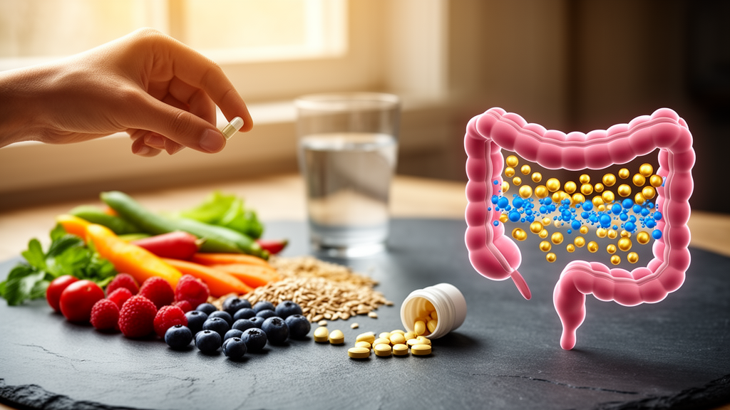
For millions of people living with Parkinson’s disease, the difference between a good day and a bad day may come down to something as ordinary as breakfast. A sweeping new review published in Frontiers in Nutrition argues that dietary habits, meal composition, and even eating behaviors such as binge eating or irregular meal timing act as powerful extrinsic modulators of anti-Parkinsonian drug performance—shaping everything from how much levodopa reaches the brain to how quickly motor symptoms return. The work, led by researchers at Shandong Medical and Pharmaceutical University and Xiamen Medical College, systematically connects phenomena that clinicians have long observed anecdotally with the molecular machinery that explains them, and then asks why this knowledge has failed to reach everyday clinical practice.
Parkinson’s disease is the second most common progressive neurodegenerative disorder worldwide, defined neuropathologically by the loss of dopaminergic neurons in the substantia nigra pars compacta and the widespread accumulation of alpha-synuclein-containing Lewy bodies. Its pharmacological backbone remains dopamine replacement therapy, anchored by levodopa—the metabolic precursor of dopamine that crosses the blood–brain barrier and is converted into the depleted transmitter within the striatum. Levodopa is still the undisputed gold standard for symptomatic control, outperforming dopamine receptor agonists, MAO-B inhibitors, COMT inhibitors, and newer non-dopaminergic agents such as the adenosine A2A receptor antagonist istradefylline in controlling bradykinetic motor features. Yet the drug’s long-term performance is notoriously fragile. Within five to ten years of continuous exposure, between 40 and 80 percent of patients develop disabling motor complications, including predictable wearing-off phenomena and levodopa-induced dyskinesias.
The review’s central technical insight is that levodopa’s fragility is baked into its pharmacokinetics. The drug has a short plasma half-life of roughly two hours, undergoes extensive peripheral metabolism, and—critically—shares a saturable transport system with dietary large neutral amino acids, or LNAAs. Levodopa relies on the L-type amino acid transporter 1, LAT1, to cross both the intestinal epithelium and the blood–brain barrier. When a protein-rich meal floods the circulation with LNAAs derived from dietary protein digestion, those amino acids competitively inhibit levodopa transport at both barriers. The result can be an abrupt termination of antiparkinsonian efficacy without any change in peripheral drug concentration: high-protein meals reduce central levodopa influx, precipitating sudden OFF states and delayed ON responses. This competitive dynamic has direct human evidence behind it. Randomized and crossover studies dating back decades have shown poorer motor response with high-protein intake and improvement with low-protein or protein-redistribution strategies in susceptible patients.
Protein competition is only one of several food-driven mechanisms the review dissects. Gastric emptying rate is the second major determinant of oral levodopa disposition. High caloric density and elevated lipid content prolong gastric emptying by inducing shallow peristalsis and enhancing antral retention, delaying the intestinal transfer of co-administered drugs. In patients receiving controlled-release levodopa/carbidopa, postprandial dosing delayed levodopa appearance and the time to peak plasma concentration, reduced the six-hour area under the curve, and delayed the onset of motor response compared with premeal administration. Meal timing therefore emerges as one of the better-supported behavioral determinants of oral levodopa response, alongside protein quantity and distribution across the day. The review also notes that non-protein constituents contribute variably: coffee, dietary fiber, and vitamin C have been observed to favorably support levodopa bioavailability, while specific botanical substances such as fresh banana juice markedly impair systemic exposure. Notably, macronutrient manipulations like the ketogenic diet show minimal effect on plasma levodopa kinetics, underscoring that competitive amino acid inhibition and gastric transit remain the predominant dietary determinants of levodopa efficacy.
Perhaps the most striking mechanism involves the gut microbiome itself. Gut bacteria expressing tyrosine decarboxylase can metabolize levodopa into peripheral dopamine before mucosal uptake, prematurely consuming the drug and drastically reducing brain delivery. The classic example is Enterococcus faecalis, whose enzymatic activity dictates local drug breakdown within the intestinal lumen. Westernized dietary patterns—characterized by excessive saturated fats, ultra-processed foods, and refined sugars—reshape gastrointestinal architecture and disrupt gut microbiota homeostasis, fostering a pro-inflammatory microbial environment that may promote bacterial levodopa metabolism. The review is careful to distinguish direct clinical evidence from mechanistic plausibility here: the proposed sequence linking Western diet-induced dysbiosis, increased bacterial levodopa degradation, and poorer therapeutic response remains largely indirect until prospective studies specifically measure medication response in Parkinson’s patients.
The microbiota–gut–brain axis, or MGBA, extends this story from drug disposition to disease progression itself. In Parkinson’s disease, pathological dietary habits trigger dysbiosis characterized by a loss of anti-inflammatory, short-chain fatty acid–producing taxa and an overgrowth of pro-inflammatory pathobionts. This microbial imbalance compromises gut mucosal barrier integrity and increases intestinal permeability, facilitating systemic dissemination of lipopolysaccharides that provoke chronic low-grade neuroinflammation, microglial activation, and blood–brain barrier dysfunction. Retrograde transport of preformed alpha-synuclein aggregates via the vagus nerve—a core neural pathway of the MGBA—directly connects gut dysregulation to progressive dopaminergic neuron loss, supporting a gut-first hypothesis in disease pathogenesis. Altered microbial metabolism also disrupts synthesis of neuroactive molecules such as gamma-aminobutyric acid, tryptophan derivatives, and secondary bile acids acting through farnesoid X receptor and TGR5 pathways, impairing central appetite and satiety signaling while exacerbating hypothalamic–pituitary–adrenal axis dysregulation.
Eating behaviors themselves add another layer of complexity. Dopaminergic therapy, particularly dopamine receptor agonists such as pramipexole and ropinirole, can precipitate impulse-control disorders that include compulsive binge eating. Large or protein-rich meals during such episodes delay gastric emptying and increase LNAA competition with levodopa, contributing to erratic absorption and unexpected motor fluctuations. Conversely, restrictive eating and unstructured calorie restriction may compromise nutritional adequacy and alter drug-metabolizing enzymes such as CYP3A4 and UDP-glucuronosyltransferases, reducing central drug efficacy even as they promote autophagy. Irregular meal timing—frequent unscheduled snacking and inconsistent daily meal times—causes fluctuating gastric pH, erratic gastric emptying, and altered splanchnic blood flow, leading to unpredictable plasma drug concentrations. The review emphasizes that medication–meal schedules should be individualized according to levodopa formulation, nutritional status, gastrointestinal function, and observed clinical response rather than applying a universal fasting strategy.
Regional dietary patterns receive a nuanced treatment that resists simple generalization. The Mediterranean diet and its MIND variant are associated with favorable neurological and metabolic outcomes—higher adherence has been linked to lower odds of Parkinson’s disease, later age at onset, and lower prodromal disease probability in longitudinal studies—yet direct evidence that these patterns improve the efficacy or pharmacokinetics of specific anti-Parkinsonian medications remains limited. Traditional East Asian dietary components show mechanistic promise: soy protein combined with time-restricted feeding suppresses MAO-B activity and preserves dopaminergic neurons in Parkinsonian models, while green tea polyphenols such as epigallocatechin-3-gallate act as multitarget agents capable of inhibiting MAO-B and preventing alpha-synuclein aggregation, though high concentrations can competitively inhibit intestinal uptake transporters such as OATP1A2 and OATP2B1. Fiber-rich traditional African dietary patterns support intestinal microbial diversity and mucosal function, and resistant starch intervention has been shown to selectively enrich beneficial taxa such as Faecalibacterium species while alleviating motor and non-motor symptoms in Parkinson’s cohorts. Legume- and maize-based Latin American diets provide high concentrations of plant proteins and LNAAs that influence systemic amino acid pools, though direct evidence that these patterns specifically alter levodopa pharmacokinetics is lacking.
The review’s most consequential argument concerns clinical translation. Despite decades of pharmacokinetic insight, the absence of precision nutrition frameworks, protein-redistribution diets, targeted chronotherapeutic meal scheduling, and multidisciplinary nutritional support substantially hinders effective translation of basic science into practice. Observational clinical data reveal that improper timing relative to meals occurs in up to 15.7 percent of general oral drug administrations. The authors advocate a paradigm shift toward precision nutrition therapy that integrates multiomics datasets—genomics, metabolomics, metagenomics—with artificial intelligence, machine learning, and smart wearable systems to predict individual metabolic responses to specific nutrient timing and food combinations. Practical interventions include administering levodopa 30 to 60 minutes before meals or two hours after, restricting protein during daytime and consuming it primarily at dinner, and establishing standardized daily meal schedules. Targeted microbiome restoration through prebiotics, probiotics, and fecal microbiota transplantation represents a promising non-invasive avenue to suppress neuroinflammation and stabilize drug pharmacokinetics. Ultimately, the review positions diet not as a peripheral lifestyle consideration but as an essential non-pharmacological pillar of Parkinson’s care—one that could stabilize drug absorption, minimize motor fluctuations, and potentially mitigate disease progression when aligned with individual drug absorption characteristics through data-driven, personalized protocols.
Subject of Research: health medicine
Article Title: Effects of eating behavior patterns and dietary habits on the therapeutic efficacy of Parkinson’s disease medications: from phenomena, mechanisms to clinical applications
Article References: Ye, J., Zhang, S. J., Guan, J., Shen, Z.-H., Wang, J., Yang, R., Huang, M., Li, Z.-M., Lin, Y., Gan, H., Wu, S., Wang, Y., & Yang, K. (2026). Effects of eating behavior patterns and dietary habits on the therapeutic efficacy of Parkinson’s disease medications: from phenomena, mechanisms to clinical applications. Frontiers in Nutrition, 13, Article 1969279. https://doi.org/10.3389/fnut.2026.1969279
Image Credits: AI Generated
DOI: 10.3389/fnut.2026.1969279
Keywords: Parkinson's disease, levodopa, nutrition, pharmacokinetics, gut microbiome, precision nutrition, LAT1 transporter, protein redistribution, Mediterranean diet, microbiota-gut-brain axis, Effects, eating
Cite Scienmag News
Diana Fleming. (October 19, 2026). What You Eat Could Decide How Well Parkinson’s Drugs Work. Scienmag. https://scienmag.com/what-you-eat-could-decide-how-well-parkinsons-drugs-work/
Diana Fleming. "What You Eat Could Decide How Well Parkinson’s Drugs Work." Scienmag, 19 October 2026, https://scienmag.com/what-you-eat-could-decide-how-well-parkinsons-drugs-work/. Accessed 19 October 2026.
Diana Fleming. "What You Eat Could Decide How Well Parkinson’s Drugs Work." Scienmag. October 19, 2026. https://scienmag.com/what-you-eat-could-decide-how-well-parkinsons-drugs-work/


