


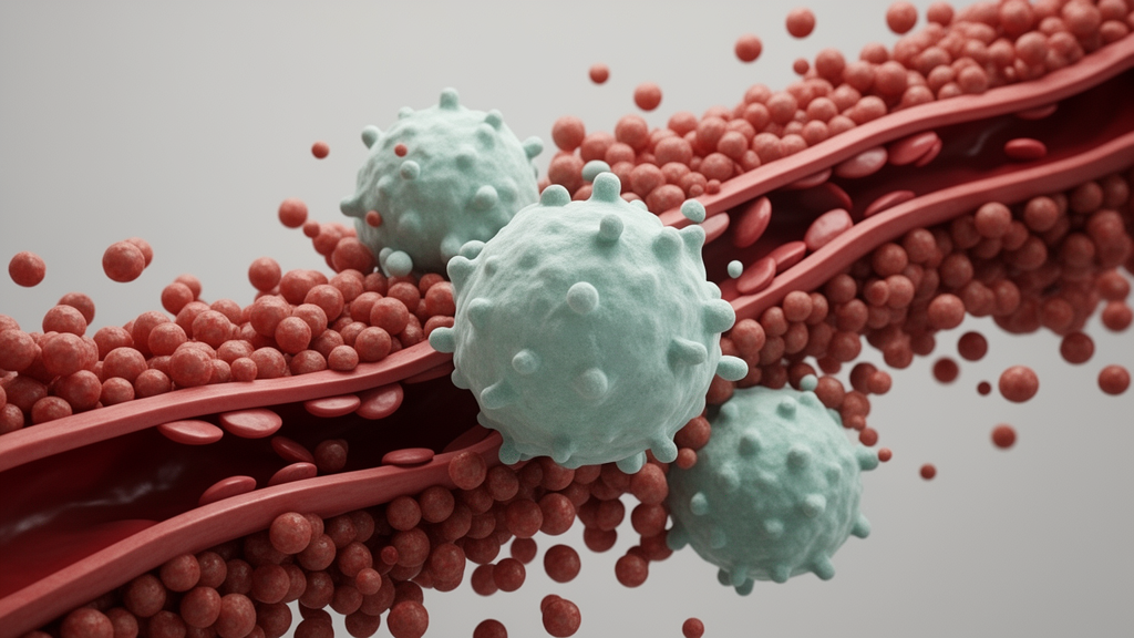
Liver cancer remains one of the most formidable challenges in oncology, and one of the chief reasons is a phenomenon known as microvascular invasion, or MVI. In hepatocellular carcinoma, the most common form of primary liver cancer, MVI refers to the presence of tumor cells within small blood vessels surrounding the tumor. Its presence is a predominant risk factor for recurrence after surgery, because it signals that malignant cells have already gained access to the vascular highway that can carry them to new sites in the liver and beyond. Now, a team of researchers at The First Affiliated Hospital of Sun Yat-sen University in Guangzhou, China, has uncovered a surprising cellular culprit that appears to help create the conditions for this vascular dissemination: a subset of immune cells that normally act as rapid responders against infection but that, under conditions of tumor stress, appear to switch roles and help build an immunosuppressive niche around the tumor.
The cells in question are mucosal-associated invariant T cells, commonly abbreviated as MAIT cells. These are unconventional T lymphocytes that recognize microbial metabolites presented by a molecule called MHC class I-related protein 1, or MR1. MAIT cells are abundant in the human liver, a location that makes them particularly relevant to hepatocellular carcinoma, and they are typically prized for their capacity to mount swift cytotoxic responses against bacteria and, potentially, against tumor cells. In the new study, published in the Journal of Translational Medicine, the researchers report that a distinct subpopulation of these cells, which they term stress-responsive MAIT cells or MAIT_SR cells, correlates strongly with the presence of microvascular invasion in patient tumors, and that these cells appear to orchestrate a peritumoral environment that favors immune escape and vascular spread.
To reach these conclusions, the team employed single-cell RNA sequencing on hepatocellular carcinoma specimens that were either positive or negative for microvascular invasion. This technique allows researchers to profile gene expression in thousands of individual cells, revealing the phenotypic landscape of the tumor microenvironment at unprecedented resolution. By integrating these single-cell data with multi-source bulk transcriptomic datasets and spatial transcriptomics, which maps gene expression onto intact tissue architecture, the investigators delineated the distinct stress-responsive phenotype of the MAIT cell population. The findings were then validated using flow cytometry, multiplex immunohistochemistry, and in vitro functional assays, providing multiple independent lines of evidence for the existence and behavior of this unusual cell subset.
What distinguishes the MAIT_SR cells from their non-stress-responsive counterparts is a striking molecular signature. The stress-responsive subset exhibited elevated expression of heat shock proteins, the molecular chaperones that cells deploy when confronted with physiological stress, but at the same time showed diminished cytotoxicity, meaning they had lost much of their tumor-killing capacity. Pathway enrichment analyses revealed that these cells were characterized by heightened activity in hypoxia signaling, glycolysis, and NF-κB signaling, all of which are hallmarks of cells adapting to the hostile, oxygen-poor, nutrient-competitive conditions that prevail within and around solid tumors. In other words, the very stressors generated by the tumor appear to reprogram these immune cells, converting potential anti-tumor allies into components of a pro-tumor ecosystem.
Perhaps the most consequential discovery concerns what these reprogrammed cells actually do. The researchers found that MAIT_SR cells were a dominant cellular source of CCL20, a chemokine, or chemical messenger, that attracts other immune cells expressing its receptor, CCR6. Using orthotopic liver cancer models, in which tumors are established in the liver of experimental animals, the team assessed the relationship between the CCL20–CCR6 axis and microvascular invasion in vivo. The results were compelling: administration of CCL20 was associated with increased MVI progression, whereas pharmacological blockade of CCR6 reduced the formation of microvascular invasion. This established the chemokine axis not merely as a correlate but as a functional driver of the invasive phenotype, at least in the experimental setting.
The mechanism by which MAIT_SR cells promote immunosuppression was further dissected through spatial analysis and functional assays. The stress-responsive MAIT cells were found to localize to peritumoral regions, the tissue immediately surrounding the tumor, where they recruited CD4-positive T cells via the CCL20–CCR6 axis. Rather than simply attracting any immune cells, however, the MAIT_SR cells actively shaped the fate of their recruits. Through the secretion of transforming growth factor beta 1, or TGF-β1, a potent immunoregulatory cytokine, they drove the differentiation of the recruited CD4-positive T cells into regulatory T cells, or Tregs. Tregs are the immune system’s brakes, cells whose physiological role is to suppress excessive immune responses but which, in cancer, are co-opted by tumors to shield malignant cells from immune attack. The study thus delineates a MAIT_SR–CCL20/TGF-β1–CD4-positive T cell axis that links stress signaling to the construction of an immunosuppressive peritumoral niche.
Beyond the mechanistic insights, the work carries potential therapeutic implications, which the researchers explored directly. In their animal models, two pharmacological interventions showed efficacy in reducing microvascular invasion: blockade of the CCR6 receptor using specific antagonists, and treatment with Acetyl-6-formylpterin, abbreviated Ac-6-FP, a compound that acts on the MR1-dependent antigen presentation pathway central to MAIT cell biology. The finding that Ac-6-FP reduced MVI formation suggests that modulating MAIT cell function through their cognate antigen-presenting molecule may be a viable strategy, while CCR6 antagonism offers a more downstream approach to sever the communication between stress-responsive MAIT cells and the CD4-positive T cells they recruit. Both strategies, if they can be translated safely to patients, would target the peritumoral niche rather than the tumor cells themselves, a conceptually distinct approach from conventional cytotoxic therapy.
The clinical significance of these findings rests on the central problem of postoperative recurrence in hepatocellular carcinoma. Even when tumors are completely resected, patients whose tumors show microvascular invasion face a substantially elevated risk of the disease returning, often within the remnant liver. Because MVI is frequently only detectable by pathological examination after surgery, clinicians currently lack effective tools to prevent vascular dissemination prospectively. If the MAIT_SR–CCL20/TGF-β1 axis proves to be a robust biomarker or a druggable pathway in human patients, it could open a window for perioperative interventions aimed at suppressing the immunosuppressive niche before tumor cells exploit it. The identification of a dominant CCL20-producing immune cell population also adds to a growing appreciation that unconventional T cells can undergo functional rewiring in tumors, a theme that has emerged repeatedly in recent cancer immunology research.
As with any translational study, important caveats apply. The association between stress-responsive MAIT cells and microvascular invasion was established through correlative analyses in human specimens combined with mechanistic work in animal models, and the efficacy of CCR6 antagonists and Acetyl-6-formylpterin was demonstrated in preclinical orthotopic models rather than in clinical trials. The authors themselves frame the findings as supporting a link between stress signaling and the peritumoral immunosuppressive niche, and as suggesting actionable therapeutic strategies, language that appropriately acknowledges the distance between mouse models and human medicine. Nevertheless, the convergence of single-cell transcriptomics, spatial mapping, flow cytometry, multiplex immunohistochemistry, and in vivo pharmacology provides an unusually comprehensive evidentiary foundation. For a disease in which vascular invasion quietly seals the fate of many surgical patients, the identification of a stress-responsive immune axis that can be pharmacologically interrupted represents a genuinely promising lead, and one that the field of hepatocellular carcinoma research will be watching closely as these findings move toward clinical validation.
Subject of Research: The role of stress-responsive MAIT cells in immunosuppression and microvascular invasion in hepatocellular carcinoma
Article Title: Stress-responsive MAIT cells are associated with immunosuppressive microenvironment and microvascular invasion in hepatocellular carcinoma
Article References: Huang, H., Xu, B., Zhang, G., Ye, D., Liao, B., Nong, Z., Guan, Y., Hu, L., Wang, X., & Li, S. (2026). Stress-responsive MAIT cells are associated with immunosuppressive microenvironment and microvascular invasion in hepatocellular carcinoma. Journal of Translational Medicine. https://doi.org/10.1186/s12967-026-09071-4
Image Credits: AI Generated
DOI: 10.1186/s12967-026-09071-4
Keywords: hepatocellular carcinoma, MAIT cells, microvascular invasion, tumor microenvironment, CCL20, CCR6, TGF-β1, regulatory T cells, single-cell RNA sequencing, immunosuppression, heat shock proteins, Acetyl-6-formylpterin
Cite Scienmag News
Nathaniel Bowman. (October 9, 2026). Stressed immune cells may help liver cancer invade blood vessels, study finds. Scienmag. https://scienmag.com/stressed-immune-cells-may-help-liver-cancer-invade-blood-vessels-study-finds/
Nathaniel Bowman. "Stressed immune cells may help liver cancer invade blood vessels, study finds." Scienmag, 9 October 2026, https://scienmag.com/stressed-immune-cells-may-help-liver-cancer-invade-blood-vessels-study-finds/. Accessed 9 October 2026.
Nathaniel Bowman. "Stressed immune cells may help liver cancer invade blood vessels, study finds." Scienmag. October 9, 2026. https://scienmag.com/stressed-immune-cells-may-help-liver-cancer-invade-blood-vessels-study-finds/


