


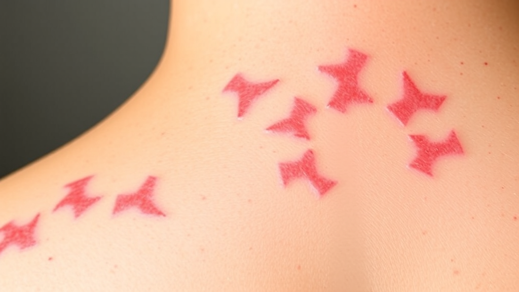
Psoriasis affects roughly two to three percent of people worldwide, producing the red, scaly, thickened skin patches that define the disease and, in many patients, paving the way for comorbidities ranging from metabolic syndrome and cardiovascular disease to depression and anxiety. Although biologic drugs that block interleukin-17 and interleukin-23 have transformed treatment for severe cases, there is still no definitive cure, and the search for cheaper, safer, topically applicable therapies continues. A new study published in the Journal of Cellular and Molecular Medicine now reveals an unexpected molecular link between vitamin D signalling and a little-studied inflammatory protein, offering a fresh explanation for why calcitriol, the active form of vitamin D, works against psoriatic skin inflammation.
The research team, led by Qingqing He and Junlin Liu, set out to map the molecular targets through which calcitriol exerts its anti-psoriatic effects. Calcitriol is best known for its role in calcium homeostasis and bone mineralization, but it also regulates cell proliferation, differentiation, oxidative stress, angiogenesis, immune modulation and antimicrobial peptide production. When calcitriol binds the vitamin D receptor, it dampens excessive T cell activation, particularly of Th17 cells, reducing interleukin-17 secretion, and it suppresses pro-inflammatory cytokines such as interleukin-6 and tumour necrosis factor-alpha. The enzyme CYP24A1 then hydroxylates calcitriol at the C-23 or C-24 position, converting it into less active metabolites that are eventually excreted, a safeguard against hypercalcaemia. What remained unclear was which downstream genes calcitriol might regulate to produce its therapeutic benefit in skin.
The researchers focused on CHI3L2, also known as YKL-39, a member of the chitinase-like protein family. Unlike its better-known relative CHI3L1, or YKL-40, CHI3L2 has been comparatively neglected in inflammatory disease research, although it is expressed in macrophages, chondrocytes and epithelial cells and can be induced by interleukin-6 and tumour necrosis factor-alpha. The protein displays chemotactic activity, growth factor activity, the ability to induce cytokine secretion and the capacity to stimulate angiogenesis. Previous work had connected CHI3L2 to the ERK1/2 signalling pathway in glioblastoma, and to STAT3 phosphorylation in invasive ductal carcinoma, where its role appears to depend on the molecular subtype of breast cancer. Because STAT3 is a critical transcription factor downstream of interleukin-6, interleukin-17, interleukin-22 and epidermal growth factor, and because persistent STAT3 activation in psoriatic keratinocytes and immune cells drives hyperproliferation, anti-apoptosis and inflammatory cytokine production, the team hypothesized that calcitriol might relieve psoriatic inflammation by suppressing CHI3L2 through inhibition of STAT3 signalling.
To test this idea in living animals, the researchers used the imiquimod-induced mouse model of psoriasis, in which topical application of the toll-like receptor 7/8 agonist activates the interleukin-23/interleukin-17 inflammatory axis that closely mirrors human disease pathogenesis. In a unilateral ear model, BALB/c mice received daily topical calcitriol solution before imiquimod cream for seven consecutive days. The treatment significantly reduced the clinical hallmarks of erythema, scaling and thickening, and histological examination showed markedly attenuated epidermal hyperplasia. Real-time quantitative PCR of ear tissue revealed substantially reduced expression of the pro-inflammatory cytokines interleukin-1alpha, interleukin-6, interleukin-17A and interleukin-23A. Importantly, serum calcium levels rose only to 2.500 millimoles per litre, comfortably within the normal physiological range of 2.0 to 2.6, and body weight, haematological parameters and organ histopathology remained unremarkable, indicating an excellent safety profile at the dose used.
The most striking finding emerged from a bilateral ear model designed to test whether local treatment could influence distant lesions. After seven days of calcitriol and imiquimod application to the right ear, the untreated left ears of the same mice were challenged with imiquimod alone for five days. Remarkably, even though only the right ear had ever received calcitriol, the left ears showed only mild psoriasiform changes compared with controls, with attenuated epidermal hyperplasia and reduced cytokine expression. Serum calcium returned to normal within five days of stopping treatment. This systemic effect suggests that calcitriol or its downstream mediators may enter the circulation and act at distal sites, although the precise circulating factors responsible remain to be identified. The observation raises intriguing questions about whether topical vitamin D therapy could benefit psoriatic lesions beyond the site of application in patients.
To dissect the cellular mechanisms, the team turned to HaCaT human keratinocytes stimulated with a five-cytokine cocktail, abbreviated M5, comprising interleukin-17A, interleukin-22, interleukin-1alpha, oncostatin M and tumour necrosis factor-alpha, which mimics the psoriatic inflammatory milieu. In psoriasis, the intermediate filament protein KRT1 is downregulated, marking impaired differentiation, while KRT6 is upregulated, indicating excessive proliferation and inflammatory activation. Calcitriol reversed both changes, restoring KRT1 and suppressing KRT6. CCK-8 and colony-formation assays showed that calcitriol counteracted the abnormal proliferation induced by M5, wound-healing assays demonstrated reduced aberrant migration, and quantitative PCR confirmed downregulation of interleukin-1alpha, interleukin-6, interleukin-17A and interleukin-23A messenger RNA.
The pivotal clue came from RNA sequencing of M5-stimulated cells treated with or without calcitriol. The analysis identified 537 differentially expressed genes, 344 upregulated and 193 downregulated. As expected, CYP24A1, the canonical calcitriol-metabolizing enzyme, was the most significantly upregulated gene, confirming that the vitamin D pathway was engaged. But the most significantly downregulated gene was CHI3L2, a result the researchers validated by quantitative PCR. When the team silenced CHI3L2 using short hairpin RNA, the effects phenocopied calcitriol treatment: KRT1 and KRT6 levels normalized, cell viability and colony formation declined, migratory capacity was impaired, and pro-inflammatory cytokine expression dropped. These loss-of-function experiments position CHI3L2 as a pro-inflammatory and pro-proliferative factor in psoriatic keratinocytes and a critical downstream effector of calcitriol.
Next, the researchers asked which signalling pathway mediates the CHI3L2 effect. KEGG pathway enrichment analysis of the downregulated genes pointed strongly to the STAT pathway, with no significant enrichment of the ERK pathway. Western blotting confirmed this: silencing CHI3L2 reduced phosphorylated STAT3 at both Tyr705 and Ser727 as well as total STAT3 protein, while phosphorylated and total ERK1/2 remained unchanged. Comparing the two STAT3 phosphorylation sites revealed that Tyr705 was the dominant regulatory node, with Ser727 showing a smaller, less pronounced change. Tyr705 phosphorylation is essential for STAT3 dimerization, nuclear translocation and transcriptional activity, so CHI3L2 appears primarily to facilitate STAT3 activation at this residue. The reduction in total STAT3 protein upon CHI3L2 knockdown also raises the possibility that CHI3L2 stabilizes STAT3 or enhances its expression, a hypothesis the authors flag for future investigation.
The authors are careful to acknowledge the limitations of their work. The imiquimod model, while widely accepted, does not fully capture the chronic, relapsing nature of human psoriasis and involves TLR7/8-driven inflammation that may not perfectly mirror the human disease. The in vitro experiments relied on HaCaT cells, an immortalized keratinocyte line, and validation in primary human keratinocytes or organotypic skin models would strengthen physiological relevance. Crucially, the study does not establish whether STAT3 or the vitamin D receptor directly binds the CHI3L2 promoter; chromatin immunoprecipitation and luciferase reporter assays would be needed to determine whether the regulation is direct or mediated through secondary transcription factors. It also remains unknown whether CHI3L2 interacts with STAT3 directly or acts through upstream receptor-mediated signalling. The systemic improvement seen in the bilateral ear model demands further exploration of circulating factors such as calcium, vitamin D metabolites or soluble inflammatory mediators.
Nevertheless, the study delivers a compelling new framework for understanding how vitamin D-based therapies work in psoriasis. By identifying CHI3L2 as a previously unrecognized downstream effector of calcitriol and demonstrating that its suppression depends on inhibiting STAT3 phosphorylation at Tyr705, the research links vitamin D signalling to chitinase-like protein regulation for the first time in cutaneous inflammation. The findings suggest that CHI3L2 could serve as a biomarker predicting response to vitamin D treatment, and that combining CHI3L2 inhibition with calcitriol administration might yield additive or synergistic benefits. With psoriasis prevalence rising worldwide and no cure in sight, uncovering a druggable node in the STAT3/CHI3L2 axis offers both mechanistic insight and a tangible target for the next generation of anti-psoriatic therapies.
Subject of Research: The mechanism by which calcitriol suppresses psoriatic inflammation and keratinocyte hyperproliferation through downregulation of CHI3L2 via the STAT3 pathway
Article Title: Downregulating CHI3L2 via the STAT3 Pathway: The Mechanism of Calcitriol in Suppressing Psoriatic Inflammation and Keratinocyte Hyperproliferation
Article References: He, Q., & Liu, J. (2026). Downregulating CHI3L2 via the STAT3 Pathway: The Mechanism of Calcitriol in Suppressing Psoriatic Inflammation and Keratinocyte Hyperproliferation. Journal of Cellular and Molecular Medicine, 30(18), Article e71367. https://doi.org/10.1111/jcmm.71367
Image Credits: AI Generated
DOI: 10.1111/jcmm.71367
Keywords: psoriasis, calcitriol, vitamin D, CHI3L2, STAT3, keratinocytes, imiquimod model, inflammation, HaCaT cells, CYP24A1, interleukin-17, drug mechanism
Cite Scienmag News
Juliet Wilcox. (September 21, 2026). Vitamin D Compound Tames Psoriasis by Switching Off a Hidden Inflammatory Gene. Scienmag. https://scienmag.com/vitamin-d-compound-tames-psoriasis-by-switching-off-a-hidden-inflammatory-gene/
Juliet Wilcox. "Vitamin D Compound Tames Psoriasis by Switching Off a Hidden Inflammatory Gene." Scienmag, 21 September 2026, https://scienmag.com/vitamin-d-compound-tames-psoriasis-by-switching-off-a-hidden-inflammatory-gene/. Accessed 5 October 2026.
Juliet Wilcox. "Vitamin D Compound Tames Psoriasis by Switching Off a Hidden Inflammatory Gene." Scienmag. September 21, 2026. https://scienmag.com/vitamin-d-compound-tames-psoriasis-by-switching-off-a-hidden-inflammatory-gene/


