


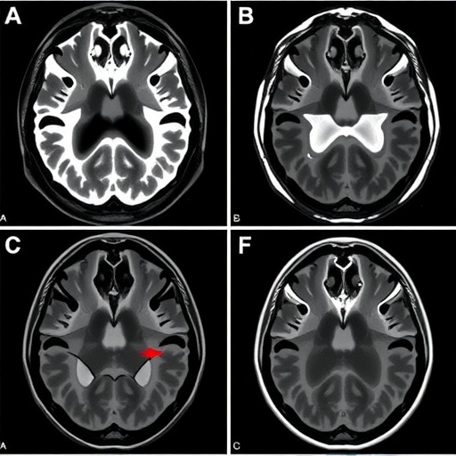
When a brain aneurysm ruptures, blood can spill into several different compartments of the skull, and where that blood ends up turns out to matter enormously for what happens next. A new study published in the journal Neurocritical Care has found that patients whose ruptured aneurysms bleed directly into the brain parenchyma—a condition known as aneurysmal intracerebral hemorrhage, or aICH—face significantly higher odds of developing cerebral vasospasm and delayed cerebral ischemia (DCI), two of the most feared delayed complications in neurocritical care. The findings come from a retrospective single-center cohort study led by Nada Babtain, Gabriel Neves, Danielle Sandsmark, and Rajat Dhar of Washington University School of Medicine in St. Louis, in collaboration with the University of Pennsylvania, and they carry direct implications for how clinicians monitor and manage patients in the days following aneurysmal rupture.
The research team analyzed 442 patients admitted with acute aneurysmal bleeding between January 2014 and November 2021 at their quaternary referral center. All patients were captured through a prospective aneurysm database, with aneurysmal rupture confirmed either by angiography or, in moribund patients who could not undergo formal angiography, presumed based on a diffuse bleeding pattern on computed tomography. Of the 442 eligible patients, 83—about 19 percent—had intracerebral hemorrhage visible on admission CT. This figure sits comfortably within the 10 to 38 percent prevalence range cited in prior literature, reinforcing that parenchymal bleeding is a common radiographic feature of aneurysmal rupture rather than a rare event. Remarkably, only eight patients, roughly 2 percent of the entire cohort, had isolated intracerebral hemorrhage without any accompanying subarachnoid hemorrhage (SAH)—a presentation so uncommon that the authors describe their analysis of it as exploratory and hypothesis-generating.
The radiographic anatomy of these hemorrhages is instructive. The median hematoma volume among patients with aICH was 12 milliliters, with the frontal lobe being the most frequent location at 61 percent of cases, followed by the temporal lobe at 46 percent. Involvement of the cerebellum and corpus callosum was rare, at just 2 percent and 8 percent respectively. Patients with aICH were older than those without it, presented with worse clinical grades on the World Federation of Neurosurgical Societies (WFNS) scale, had thicker cisternal blood on the modified Fisher scale, higher intraventricular hemorrhage scores, and paradoxically smaller ruptured aneurysms. One of the most striking associations was aneurysm location: while anterior cerebral artery and anterior communicating artery aneurysms were the most common in both groups, patients with aICH were far more likely to have ruptured middle cerebral artery (MCA) aneurysms—24 percent versus 8 percent in those without ICH, a difference that reached statistical significance at p = 0.0001. This aligns with a broader literature suggesting that MCA aneurysms, by virtue of their proximity to the sylvian fissure and brain parenchyma, are particularly prone to producing parenchymal hematomas when they burst.
To assess the delayed complications, the investigators focused on the 356 patients—81 percent of the cohort—who survived at least four days after hemorrhage. This cutoff reflects the clinical reality that vasospasm and DCI typically begin to manifest around day four, meaning that deaths occurring before this window represent a fundamentally different failure mode rather than a failure to develop these endpoints. Eighty-six patients did not survive beyond 96 hours, including 24 with aICH, and these early deaths were concentrated among patients with higher clinical and radiographic severity. Among survivors, the contrast between those with and without aICH was stark: 59 percent of patients with intracerebral hemorrhage developed angiographic vasospasm compared with 34 percent of those without, and 42 percent developed delayed cerebral ischemia compared with 21 percent, both differences highly significant at p = 0.0005.
The statistical rigor applied here deserves attention. Because patients with aICH differed from those without it in age, clinical grade, and radiographic severity, the researchers used multivariable logistic regression to disentangle the independent contribution of ICH from these confounders. After adjusting for age, aneurysm size, WFNS grade, modified Fisher score, and intraventricular hemorrhage score, the presence of ICH remained an independent risk factor for vasospasm, with an odds ratio of 2.26 and a 95 percent confidence interval of 1.13 to 4.52. For DCI, the adjusted odds ratio was 1.83 with a confidence interval of 0.90 to 3.68—a trend that did not reach conventional statistical significance. Adding ICH as a variable measurably improved the vasospasm prediction model, reducing the Akaike Information Criterion from 386.8 to 381.1, with a likelihood ratio test p-value of 0.005. For DCI, the improvement was marginal and not significant. Hematoma volume itself also mattered: among patients with aICH, those who went on to develop vasospasm or DCI had larger hematomas on average, 13 milliliters versus 8 milliliters, with p-values of 0.03 and 0.04 respectively. To guard against survival bias, the team performed a worst-case sensitivity analysis in which all patients who died early were assumed to have developed both vasospasm and DCI. The association between ICH and these outcomes held, strengthening confidence in the robustness of the conclusions.
The clinical consequences of parenchymal bleeding extended beyond the delayed complications themselves. Patients with aICH spent longer in the intensive care unit and in the hospital overall, and their discharge outcomes were considerably worse. Only 7 percent of patients with aICH were discharged with a modified Rankin Scale score of 0 to 2, indicating functional independence, compared with 27 percent of those without ICH—a difference that was highly significant. Similarly, 48 percent of patients with aICH were discharged home or to a rehabilitation facility versus 72 percent of those without. Mortality was also higher in the ICH group, at 35 percent versus 21 percent. However, when the researchers adjusted for the differential admission severity, as captured by WFNS grade, these outcome differences largely disappeared. In other words, the poorer outcomes among patients with aICH appear to be driven primarily by the fact that they arrive sicker, not by the parenchymal hemorrhage itself acting as an independent arbiter of long-term disability.
The rarest and most intriguing subgroup—patients with aneurysmal rupture causing intracerebral hemorrhage without any subarachnoid blood—numbered just eight in this cohort, representing 2 percent of all aneurysmal hemorrhages and 10 percent of those with aICH. All eight had confirmed aneurysms on angiography, and half were due to ruptured MCA aneurysms. Prior published data on this presentation are sparse: a single case series reported nine patients, and a systematic review identified only 22 patients in the literature, with an estimated incidence of approximately 1.6 percent, broadly consistent with the current findings. The aneurysms in the current cohort were smaller overall, with a median size of 7 millimeters, compared with a broader range of 3 to 40 millimeters reported in prior studies. Among these eight patients, the rates of vasospasm and DCI were numerically lower than in patients with concomitant ICH and SAH—43 percent versus 62 percent for vasospasm, and 14 percent versus 46 percent for DCI—but the sample size was far too small to establish statistical significance, and the authors explicitly refrain from drawing firm conclusions about this subgroup.
The mechanistic explanation for why parenchymal blood might drive vasospasm remains speculative but biologically plausible. The leading hypothesis centers on oxyhemoglobin, a breakdown product released when red blood cells lyse. In classic aneurysmal subarachnoid hemorrhage, oxyhemoglobin accumulates in the subarachnoid spaces and triggers constriction of the major cerebral arteries, in part by scavenging nitric oxide, the endothelium-derived molecule that normally maintains vascular relaxation. The authors propose that when blood infiltrates the brain parenchyma, it can also enter the perivascular spaces—known as Virchow–Robin spaces—which are fluid-filled channels surrounding the small perforating vessels as they penetrate from the subarachnoid space into the brain tissue. Oxyhemoglobin accumulating in these spaces could promote vasoconstriction of the deep penetrating arteries through the same nitric oxide scavenging mechanism, compounding the ischemic threat posed by the mass effect of the hematoma itself. When cerebral perfusion fails to match metabolic demand—whether from large-vessel vasospasm, small-vessel constriction, or direct compression by the hematoma—delayed cerebral ischemia and infarction follow.
The practical implications of this study are immediately actionable for neurocritical care teams. Patients with aneurysmal rupture and concomitant intracerebral hemorrhage should be recognized as a high-risk group warranting heightened surveillance for vasospasm and DCI, beyond what their clinical grade alone might suggest. Even for the rare patients presenting with isolated parenchymal hemorrhage without subarachnoid blood, the study suggests that the risk of delayed complications is low but not absent—nearly half of these patients developed vasospasm—and the authors suggest it may still be reasonable to administer prophylactic nimodipine, maintain euvolemia, and continue close monitoring for DCI, even in the absence of cisternal blood on initial imaging. The study does have limitations, including its retrospective single-center design and the subjective elements inherent in DCI adjudication, though the researchers mitigated this by requiring careful evaluation of the time course and pattern of neurological deficits and by confirming vasospasm with conventional digital subtraction angiography in most positive cases. Referral bias was also considered, but the inclusion of both direct emergency department admissions and inter-hospital transfers across the full spectrum of disease severity likely enhances the representativeness of the cohort. For a condition as devastating as aneurysmal rupture, where secondary injury in the days after the initial bleed often determines the difference between recovery and permanent disability, refining the risk stratification tools available to clinicians is a meaningful step forward.
Cite Scienmag News
Cassandra Pierce. (September 3, 2026). Vasospasm and Delayed Ischemia After Aneurysmal Rupture With Hemorrhage. Scienmag. https://scienmag.com/vasospasm-and-delayed-ischemia-after-aneurysmal-rupture-with-hemorrhage/
Cassandra Pierce. "Vasospasm and Delayed Ischemia After Aneurysmal Rupture With Hemorrhage." Scienmag, 3 September 2026, https://scienmag.com/vasospasm-and-delayed-ischemia-after-aneurysmal-rupture-with-hemorrhage/. Accessed 3 September 2026.
Cassandra Pierce. "Vasospasm and Delayed Ischemia After Aneurysmal Rupture With Hemorrhage." Scienmag. September 3, 2026. https://scienmag.com/vasospasm-and-delayed-ischemia-after-aneurysmal-rupture-with-hemorrhage/


