


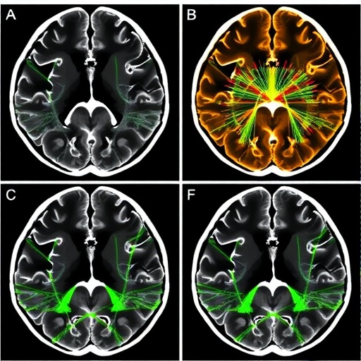
The brain’s white matter has long been described through profiles: numbers that rise and fall along the length of each major fiber bundle, summarizing the health of the cabling that connects one cortical region to another. A new systematic review published in the journal Neuroinformatics argues that this familiar one-dimensional picture may be quietly discarding some of the most clinically meaningful information in diffusion MRI. Led by Junhao Li and Ye Wu of Nanjing University of Science and Technology, together with colleagues at Shanghai Mental Health Center and Union Hospital of Huazhong University of Science and Technology, the review maps the rapidly growing field of graph-based white matter tractometry, in which fiber bundles are represented not as lines with attached measurements but as networks, or graphs, that preserve their spatial topology. According to the authors, this shift could allow researchers to detect distributed patterns of pathology that traditional along-tract analyses collapse into averages, but the field now stands at a decisive crossroads: methods are multiplying faster than the validation infrastructure needed to prove them trustworthy.
The technical foundation of the approach lies in how white matter is measured in the first place. Diffusion MRI does not image tissue microstructure directly; it measures the behavior of water molecules as they diffuse through brain tissue, and quantities such as fractional anisotropy, diffusivity, neurite orientation dispersion, and parameters from models like NODDI and diffusional kurtosis imaging are inferred from that water signal. The review emphasizes that this distinction is not pedantic. Every diffusion metric depends on model assumptions about what is happening inside voxels—how many compartments exist, how water exchanges between them, what the cells look like—and those assumptions have been validated only in restricted experimental contexts, often through histological comparisons in animals, post-mortem tissue, or physical phantoms. Interpreting a drop in fractional anisotropy as demyelination, or a change in axial diffusivity as axonal injury, carries well-documented pitfalls. Graph-based tractometry inherits all of these uncertainties from the measurement layer beneath it, which means that no amount of clever network mathematics can fully rescue an ambiguous diffusion signal.
On top of the diffusion measurements sits tractography, the computational procedure that reconstructs fiber pathways from the orientation information in the images. The review is candid about the known limitations of this spatial scaffolding. Large-scale evaluations, including the 2017 challenge of mapping the human connectome based on diffusion tractography, revealed that tractography algorithms can produce false positives and anatomically implausible streamlines, particularly in regions where fibers cross, kiss, or fan. Validation efforts such as the Tractometer, the ISMRM 2015 Tractography Challenge scoring system, and the more recent IronTract challenge have pushed the field toward more rigorous benchmarking, but the review notes that graph-based tractometry adds yet another layer of choices on top of tractography, each of which can reshape the final result.
That added layer is graph construction itself, and it is where the authors identify one of the most serious weaknesses in the literature. To turn a bundle of streamlines into a graph, researchers must decide what the nodes are and what the edges represent. Nodes might be segments along a tract, cross-sectional slices, clusters of streamlines with similar shapes, or points sampled in three-dimensional space. Edges might encode spatial adjacency, similarity of diffusion profiles, geometric distance, or some learned relationship. Each of these choices implicitly encodes a hypothesis about how pathology is organized—whether disease spreads along connected segments, whether abnormality appears in spatially coherent clusters, whether distant parts of a tract deteriorate in tandem. Yet the review finds that graph-construction decisions are frequently underspecified in published studies, described in too little detail to be reproduced and rarely tested against alternatives. Because different constructions can yield different networks from the same underlying data, the risk is that some reported findings reflect modeling choices rather than biology.
Once a graph is built, detection models take over. Here the review documents a striking convergence with the broader explosion of graph neural networks in network neuroscience. Architectures adapted from graph convolutional networks, graph attention networks, and hybrid graph-CNN-transformer designs—such as TractGraphFormer, which the review cites as an example of anatomically informed deep learning applied to tractography—can in principle learn which topological patterns distinguish patients from controls. The authors survey applications across neurological and psychiatric conditions, noting that clinical studies have demonstrated consistent group differences between patient populations and healthy participants across a range of disorders, from multiple sclerosis and gliomas to psychiatric illness. Where controlled comparisons have been performed, graph-based methods show modest improvements over traditional approaches, suggesting genuine added value rather than wholesale transformation.
The comparison targets themselves deserve explanation, because they define the standard the new methods must beat. Tract-based spatial statistics, or TBSS, introduced in 2006, remains the workhorse of group-level white matter analysis: it projects diffusion metrics onto a skeleton of the white matter and compares voxels across subjects, sidestepping some alignment problems while introducing others related to skeleton representation and non-linear registration. Automated fiber quantification, or AFQ, takes the opposite approach, extracting specific tracts and computing profiles of diffusion metrics along their length, a tradition the review traces through along-tract statistics and bundle analytics frameworks. Both approaches are mature, widely deployed, and supported by extensive reliability studies. The uncomfortable finding of the review is that comprehensive benchmarking of graph-based tractometry against TBSS and AFQ is simply absent. Studies compare new graph methods against their own ablations or against no baseline at all, leaving the community unable to judge how much of the reported improvement is real and how much is an artifact of different pipelines.
The validation gap extends beyond missing head-to-head comparisons. The review documents concrete structural barriers to systematic validation of tractometry detection models, and one of the most consequential is the absence of dedicated validation platforms. Tractography, by contrast, has benefited from nearly a decade of community infrastructure: phantom-based evaluations, challenge scoring systems, and curated reference datasets that let researchers quantify exactly how well an algorithm recovers known anatomy. Nothing comparable exists for graph-based tractometry detection. There are no standardized benchmarks against which a new graph neural network can be scored, no agreed-upon ground truth for what a graph representation of a diseased tract should look like, and no shared testing environment in which methods from different laboratories can be evaluated under identical conditions. The authors assess emerging infrastructure that could begin to address these gaps, including large curated tractometry resources built on datasets such as the Human Connectome Project and software ecosystems that standardize processing pipelines, but they conclude that dedicated validation platforms for detection models do not yet exist.
Reliability poses a further challenge that the review treats in detail. White matter varies substantially across individuals, across the lifespan, and across regions of the brain, and recent work cited in the review warns that population averages may not represent any single individual well. Multi-site imaging introduces scanner-related variability that can swamp subtle biological signals, motivating harmonization frameworks built on bundle analytics to align measurements across acquisition platforms. Tract dissection reproducibility—the extent to which different pipelines extract the same bundles from the same data—remains a persistent concern, as documented by the Tractostorm initiative. Any graph-based analysis inherits these sources of variability, and a graph constructed from an unstable tractography result will be unstable in turn, no matter how sophisticated the downstream model.
Despite these caveats, the review’s overall assessment is that graph-based tractometry shows genuine promise as a research tool. The core insight—preserving the spatial topology of fiber bundles rather than collapsing it into one-dimensional profiles—is scientifically sound and biologically motivated, because white matter pathology in many diseases is distributed and structured rather than uniform. The consistency of group differences reported across clinical studies suggests that the representations capture something real. The problem is one of maturity, not of principle. Traditional approaches reached clinical relevance only after years of reliability testing, benchmarking, and standardization, and the review argues that graph-based methods must undergo an equivalent maturation process before their findings can be translated into clinical decision support.
The path forward, as the authors sketch it, involves several concrete steps. Graph-construction choices must be specified, shared, and stress-tested as rigorously as the models built on top of them. Head-to-head benchmarking against TBSS and AFQ on common datasets should become a baseline expectation for new methods. Validation platforms analogous to the Tractometer—purpose-built for tractometry detection models rather than for tractography alone—would give the field the same feedback loop that matured its predecessor. And the biological interpretation of the diffusion metrics feeding these graphs needs continued validation against histology and phantoms, so that network-level findings rest on measurements whose microstructural meaning is understood. Until then, the review concludes, graph-based white matter tractometry should be regarded as a powerful and evolving research instrument whose clinical potential remains real but unrealized, waiting on a validation effort equal in ambition to the modeling creativity that has driven the field’s rapid growth.
Cite Scienmag News
Cassandra Pierce. (September 3, 2026). Graph-Based White Matter Tractometry: Methods, Applications, and Validation Paths. Scienmag. https://scienmag.com/graph-based-white-matter-tractometry-methods-applications-and-validation-paths/
Cassandra Pierce. "Graph-Based White Matter Tractometry: Methods, Applications, and Validation Paths." Scienmag, 3 September 2026, https://scienmag.com/graph-based-white-matter-tractometry-methods-applications-and-validation-paths/. Accessed 3 September 2026.
Cassandra Pierce. "Graph-Based White Matter Tractometry: Methods, Applications, and Validation Paths." Scienmag. September 3, 2026. https://scienmag.com/graph-based-white-matter-tractometry-methods-applications-and-validation-paths/


