


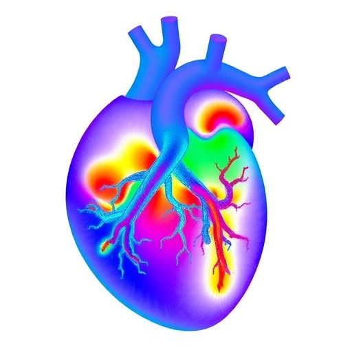
In a groundbreaking development poised to revolutionize biomedical imaging, scientists have introduced a multi-lens ultrasound array system capable of delivering unprecedented three-dimensional visualization of micro-vascular structures within entire organs. This innovative technology overcomes previous limitations in volumetric imaging resolution and scale, providing researchers and clinicians with an exceptional tool to explore the intricate network of blood vessels that sustain organ function and health.
At the heart of this advancement lies the synergistic combination of ultrasound technology with novel multi-lens architecture, enabling comprehensive capture of vascular details at a microscopic level throughout large biological specimens. Traditional ultrasound approaches have historically struggled to balance image resolution with penetration depth and scanning volume, often leaving the micro-vascularized components inadequately characterized. The multi-lens arrays bypass these constraints by leveraging a distributed arrangement of ultrasound detectors, each acting as a highly sensitive focal element that collectively reconstructs volumetric data with remarkable clarity.
The engineering design cleverly arranges multiple ultrasound lenses in an array format, dramatically expanding the imaging field of view while maintaining fine spatial resolution. This array configuration allows simultaneous acquisition of acoustic signals from different angles, which can then be computationally synthesized into a cohesive three-dimensional representation. The capacity to scan entire organs, rather than sections or superficial layers, represents an enormous stride toward comprehensive vascular mapping in vivo.
Such innovation is made possible through advancements in signal processing algorithms tailored to the unique data collected by the multi-lens system. Leveraging powerful reconstruction techniques, researchers translate raw ultrasound echoes into high-fidelity volumetric images that faithfully depict complex branching and intertwined micro-vasculature. This approach surpasses conventional single-lens delay-and-sum methods, offering enhanced signal-to-noise ratio, improved contrast, and reduced artifacts.
The implications of this imaging capability reach far into medical science and translational research. Microvascular health plays a critical role in organ function, disease progression, and response to therapies. Conditions such as cancer, diabetes, and neurodegenerative diseases often involve subtle changes in microvascular architecture that elude detection with existing clinical imaging tools. The ability to monitor and quantify these alterations non-invasively across whole organs opens new avenues for early diagnosis, therapeutic monitoring, and personalized medicine.
In addition to clinical applications, the multi-lens ultrasound arrays present unique opportunities in basic biological research. For example, developmental biologists can now dynamically visualize angiogenesis—the formation of new blood vessels—with exceptional detail, gaining insights into organogenesis and tissue regeneration. Similarly, pharmacologists can screen the vascular effects of novel drug candidates, enabling more precise assessment of safety and efficacy profiles.
The system’s versatility is further enhanced by its compatibility with conventional ultrasound equipment and ease of integration into existing imaging workflows. By utilizing widely available ultrasound transducers combined with custom multi-lens attachments and advanced computational backends, this technology balances accessibility with cutting-edge performance. This practical aspect facilitates rapid adoption across research institutions and healthcare facilities.
Moreover, the multi-lens array approach mitigates common limitations associated with ultrasound imaging, such as acoustic attenuation and scattering, especially in heterogeneous tissues. Carefully engineered lens geometries and optimized frequency ranges achieve deeper penetration depths while preserving fine vascular details. This robust performance across diverse organ types, from dense organs like the liver to delicate structures such as the brain, exemplifies the system’s adaptability.
The potential of this technology to transform diagnostic algorithms is immense. For cardiologists, detailed mapping of coronary microvasculature may allow better stratification of ischemic risks. Neurologists could benefit from real-time observation of cerebral blood flow changes linked to stroke or neurodegeneration. Oncologists might identify tumor angiogenesis signatures earlier, guiding targeted interventions. These clinical enhancements promise improved patient outcomes through more informed decision-making.
From a technical perspective, future iterations aim to further miniaturize lens components, increase array element density, and enhance real-time processing capabilities. Integrating machine learning frameworks with volumetric ultrasound data is also envisioned, facilitating automated segmentation, anomaly detection, and predictive modeling. Such integrations would exponentially boost the technology’s utility and diagnostic power.
The research team’s work opens an exciting frontier in ultrasound imaging, transforming it from a primarily planar or sectional modality into an immersive three-dimensional exploration platform. This shift aligns with broader trends toward multidimensional biomedical imaging, where the spatial complexity of biological systems is faithfully captured and analyzed. The multi-lens ultrasound system exemplifies this evolution by offering a scalable, detailed, and practical solution to a long-standing imaging challenge.
As this technology progresses toward clinical translation, ongoing collaborations between engineers, clinicians, and biologists will be essential. Rigorous validation studies, regulatory approvals, and development of new clinical protocols must accompany further technical refinements. Nevertheless, the foundational capabilities demonstrated already mark a paradigm shift in how we visualize microvascular networks and understand their roles in health and disease.
In parallel, the approach highlights the significance of interdisciplinary innovation combining physical acoustics, optical design principles adapted to ultrasound, and computational imaging. By bridging these domains, the multi-lens array ultrasound system not only advances vascular imaging but also inspires similar cross-disciplinary solutions for other biomedical challenges.
The potential social impact of this technology extends to global health, where affordable, non-invasive, and high-resolution imaging is much needed, especially in resource-limited settings. Portable ultrasound systems enhanced with multi-lens arrays could provide detailed diagnostics at the point of care, reducing dependency on costly and less accessible imaging modalities like MRI or CT scans.
Ultimately, this breakthrough heralds a new era for vascular medicine and biomedical imaging at large. By unlocking comprehensive three-dimensional views of microvascular arrangements across whole organs, the multi-lens ultrasound arrays hold the promise to deepen our biological understanding, refine disease management, and catalyze innovative therapies. The ripple effects of this advancement will be felt across scientific disciplines and clinical fields for years to come.
Cite Scienmag News
Ophelia Keating. (October 28, 2025). Multi-Lens Ultrasound Maps 3D Organ Microvasculature. Scienmag. https://scienmag.com/multi-lens-ultrasound-maps-3d-organ-microvasculature/
Ophelia Keating. "Multi-Lens Ultrasound Maps 3D Organ Microvasculature." Scienmag, 28 October 2025, https://scienmag.com/multi-lens-ultrasound-maps-3d-organ-microvasculature/. Accessed 3 September 2026.
Ophelia Keating. "Multi-Lens Ultrasound Maps 3D Organ Microvasculature." Scienmag. October 28, 2025. https://scienmag.com/multi-lens-ultrasound-maps-3d-organ-microvasculature/


