
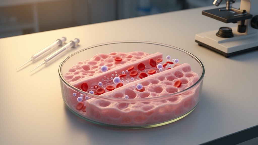
Multiple myeloma remains largely incurable, with most patients eventually relapsing or developing refractory disease, and a growing body of work argues that the reason lies partly outside the tumor cell itself. A new review published in Frontiers in Cell and Developmental Biology examines how three-dimensional laboratory models of the bone marrow microenvironment are being built to capture the biology that conventional two-dimensional cultures and animal models miss. Written by researchers at the University of Parma, the IRCCS Istituto Ortopedico Rizzoli and the Politecnico di Milano, the review offers a critical, mechanism-oriented assessment of both bone-specific and bone marrow–like 3D platforms, evaluating what each can reproduce, what readouts it enables, and where it fits in translational research.
The authors frame multiple myeloma as a malignancy that is strongly dependent on its surroundings. The disease is characterized by the clonal expansion of antibody-secreting plasma cells in the bone marrow, and progression from premalignant stages such as monoclonal gammopathy of undetermined significance and smoldering myeloma to active disease is driven not only by genetic and epigenetic lesions within the plasma cells but also by progressive remodeling of the bone microenvironment. That niche is described as a dynamic, continuously remodeled compartment in which osteoblasts, osteoclasts, osteocytes, osteogenic progenitors and the mineralized extracellular matrix interact with malignant plasma cells, disrupting bone homeostasis, promoting myeloma bone disease and contributing to therapeutic resistance.
Conventional preclinical models each carry limitations that constrain their ability to reproduce these microenvironment-dependent mechanisms. Two-dimensional cultures, including monocultures of myeloma cell lines such as RPMI-8226, U-266, MM1.S and OPM-2, are inexpensive, reproducible and compatible with high-throughput drug screening, but they largely exclude the bone marrow niche that is essential for malignant plasma cell survival, homing and drug resistance. Even two-dimensional co-cultures with stromal cells, which have shown that stromal contact and soluble factors can protect myeloma cells from therapy-induced apoptosis, remain reductionist: they fail to reproduce bone marrow architecture, physiological extracellular matrix properties, mineralized bone matrix, oxygen and nutrient gradients, and the spatial organization of stromal, bone, vascular and immune compartments. As a consequence, the review notes, they may overestimate drug sensitivity and systematically underestimate microenvironment-mediated resistance.
Animal models span a wide range of systems, each with specific strengths and constraints. Syngeneic immunocompetent models, primarily the 5TMM series, support tumor growth with an intact murine immune system and have been valuable for studying bone disease, angiogenesis and immune regulation, with the 5TGM1 model widely used because it reproduces bone marrow localization, monoclonal protein production, osteolytic lesions and altered bone remodeling. Xenograft models allow in vivo assessment of human tumor growth and drug activity but depend on immunodeficient hosts, limiting the study of adaptive immunity and immunotherapy response. Humanized bone models such as SCID-hu and SCID-rab better support myeloma growth within a human bone compartment, yet they remain technically demanding, expensive, low-throughput and dependent on immunodeficient hosts. The review concludes that no animal model fully recapitulates the human myeloma bone microenvironment.
Against this backdrop, the authors organize 3D models into several classes. Scaffold-free systems, including spheroids, organoids and multicellular aggregates, differ in their degree of cellular organization and complexity. One high-throughput spheroid co-culture system using primary myeloma cells with or without mesenchymal stromal cells supported sustained proliferation for up to 21 days and showed that bortezomib and auranofin induced lower cytotoxicity in 3D co-culture than in 3D monoculture or 2D conditions. More recently, a combined bone and lympho-myeloid organoid model supported the engraftment and survival of primary myeloma cells ex vivo for up to two weeks, with single-cell RNA sequencing confirming the low proliferative rate characteristic of myeloma and the expression of canonical markers including BCL2, IRF4 and SLAMF7.
Scaffold-based and extracellular matrix–mimetic models form a second class, built from natural polymers such as hyaluronic acid, gelatin, collagen and alginate or synthetic polymers such as polycaprolactone and polyethylene glycol. Among the notable examples, a Matrigel-based model supported expansion of myeloma clones up to 17-fold, including cells harboring clonotypic IgH VDJ rearrangements, and showed that melphalan mainly targeted hematopoietic cells whereas bortezomib selectively targeted CD138-positive CD56-positive plasma cells. A synthetic PuraMatrix hydrogel model co-culturing bone marrow mononuclear cells from patients with stromal cells reproduced resistance to bortezomib and doxorubicin reflecting clinical resistance patterns. A polycaprolactone-based polyHIPE scaffold mimicking the osteoblastic bone niche, where myeloma dormancy may occur, facilitated osteoblast and myeloma cell infiltration and altered cellular sensitivity to melphalan, bortezomib and lenalidomide. The review also highlights scaffold stiffness as a promising but underexplored parameter in the myeloma niche.
Bone-like and patient-derived tissue-engineered models add bone-specific features such as mineralized matrix, osteogenic maturation or osteoclast activity, and the authors stress that the presence of bone-associated cells alone is not sufficient to classify a model as bone-like. Decellularized bone matrices preserve native extracellular matrix composition and biomechanical cues, though the decellularization process limits reproducibility and tunability. Patient-derived models are particularly attractive for precision medicine: one system embedding bone marrow accessory and cancer cells in a fibrinogen-based matrix retrospectively predicted clinical responses to regimens including bortezomib, doxorubicin and carfilzomib in 89 percent of the myeloma cohort studied, although the authors note it was a pilot study requiring prospective validation.
Bioprinted and microfluidic platforms represent the most technologically advanced end of the spectrum. Bioprinting allows precise control of cellular distribution, matrix composition and tissue architecture, enabling compartmentalized microenvironments that mimic endosteal and perivascular niches. One model combined bioprinted calcium phosphate cement scaffolds mimicking the mineralized endosteal niche with a Matrigel-based perivascular compartment containing stromal cells and endothelial progenitor cells, supporting the survival and localization of primary CD138-positive plasma cells. A coaxially bioprinted model with a nano-hydroxyapatite-enriched outer sheath promoted myeloma cell proliferation, enhanced IL-6 secretion and reproduced stromal-mediated resistance to bortezomib; notably, inhibition of IL-6 signaling with tocilizumab restored sensitivity to bortezomib, illustrating how such systems can identify microenvironment-dependent therapeutic vulnerabilities. Vascularized myeloma-on-chip models integrating endosteal and perivascular niches with perfusable microvascular networks have supported long-term survival of both cell lines and patient-derived plasma cells and enabled dynamic delivery of anti-BCMA CAR-T cells with real-time assessment of immune-cell trafficking and tumor cytotoxicity.
The review emphasizes that these models should be considered complementary rather than a linear progression in which increasing complexity necessarily yields greater physiological relevance. Simpler scaffold-free and hydrogel-based models offer more accessible, controllable workflows, whereas multicellular tissue-engineered, bioprinted and microfluidic systems reproduce a broader range of microenvironmental features at the cost of more complex fabrication and analysis. Patient-derived material increases biological relevance but is influenced by sample availability, quality and interpatient heterogeneity, affecting reproducibility and scalability. Model selection, the authors argue, should be driven by the biological question, the required readout and practical constraints rather than by overall complexity.
On the translational front, the authors identify patient-derived 3D cultures as promising functional platforms for ex vivo drug testing that could complement molecular profiling, particularly in relapsed or refractory myeloma where clonal evolution and microenvironment-mediated resistance complicate therapeutic selection. However, they caution that biological relevance should not be equated with clinical predictivity: most 3D models have been validated against experimental or molecular endpoints, and direct comparisons between ex vivo drug responses and clinical outcomes remain uncommon and have generally involved limited patient cohorts. Prospective validation in larger, independent populations is needed before such assays can be considered clinically predictive. The review also examines computational and artificial intelligence approaches, noting that while deep-learning pipelines have mapped spatial heterogeneity of malignant plasma cells in patient biopsies, direct application of AI to 3D myeloma models remains limited and faces challenges including small datasets, batch variability and overfitting risk. The authors conclude that 3D models are likely to contribute as complementary systems for mechanistic investigation and translational research rather than replacing established preclinical approaches, with future progress depending on modular, standardized platforms designed around defined biological questions.
Subject of Research: Three-dimensional models of the multiple myeloma bone microenvironment and their translational applications
Article Title: Three-dimensional models of the multiple myeloma bone microenvironment: from disease mechanisms to translational applications
Article References: Sitzia, C., Manferdini, C., D’Atri, G., Lisignoli, G., Dessena, M., Giuliani, N., & Storti, P. (2026). Three-dimensional models of the multiple myeloma bone microenvironment: from disease mechanisms to translational applications. Frontiers in Cell and Developmental Biology, 14, 1954766. https://doi.org/10.3389/fcell.2026.1954766
Image Credits: AI Generated
DOI: 10.3389/fcell.2026.1954766
Keywords: multiple myeloma, bone microenvironment, 3D models, organoids, bioprinting, organ-on-chip, drug resistance, myeloma bone disease, tissue engineering, patient-derived models, bone remodeling, precision medicine
Cite Scienmag News
Gregory Coleman. (October 29, 2026). Lab-Grown Bone Marrow Models Reshape How Myeloma Is Studied. Scienmag. https://scienmag.com/lab-grown-bone-marrow-models-reshape-how-myeloma-is-studied/
Gregory Coleman. "Lab-Grown Bone Marrow Models Reshape How Myeloma Is Studied." Scienmag, 29 October 2026, https://scienmag.com/lab-grown-bone-marrow-models-reshape-how-myeloma-is-studied/. Accessed 29 October 2026.
Gregory Coleman. "Lab-Grown Bone Marrow Models Reshape How Myeloma Is Studied." Scienmag. October 29, 2026. https://scienmag.com/lab-grown-bone-marrow-models-reshape-how-myeloma-is-studied/