


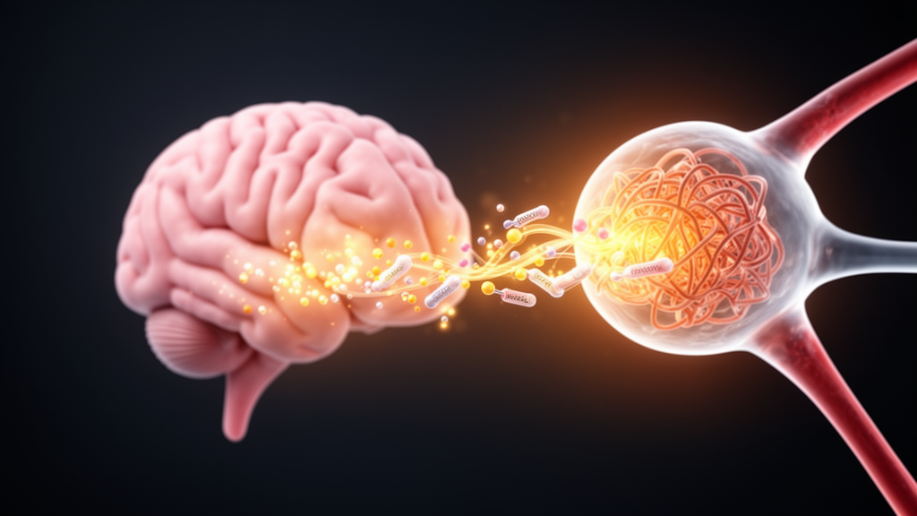
When a newborn’s brain is starved of oxygen and blood during birth, the damage does not stop when oxygen returns. In the hours and days that follow, a hidden cascade of cellular failure unfolds inside neurons, and clinicians currently have only one weapon against it: therapeutic hypothermia, or controlled cooling, which helps many infants but leaves others with cerebral palsy, epilepsy, or lifelong cognitive impairment. Now a comprehensive review published in Frontiers in Molecular Neuroscience argues that an unexpected class of molecules, produced not by the brain but by the bacteria living in the gut, could offer a fundamentally new way to protect the newborn brain. The molecules are short-chain fatty acids, and the review’s central claim is striking: they may work by dismantling a self-amplifying vicious cycle between two of the cell’s most important organelles.
The review, led by Yinyu Wang and Zhidan Bao of Jiangyin People’s Hospital together with colleagues at Jiangyin Clinical College of Xuzhou Medical University and Wuxi Taihu University, focuses on neonatal hypoxic-ischemic encephalopathy, or HIE, a devastating condition caused by perinatal asphyxia and one of the leading causes of neonatal death and disability worldwide. Therapeutic hypothermia remains the standard of care, but its benefits are most reliable in mild cases, its treatment window is narrow, and eligibility criteria exclude many infants who might benefit. The authors argue that the persistence of severe sequelae in cooled infants demands adjunctive therapies aimed at mechanisms that cooling does not address, and they identify the gut-brain axis as a promising frontier.
Short-chain fatty acids, chiefly acetate, propionate, and butyrate, are saturated fatty acids of one to six carbons that gut bacteria generate by fermenting dietary fiber in the colon. Together these three metabolites account for more than ninety percent of total short-chain fatty acids in the body. Butyrate serves as the primary energy source for colonic epithelial cells, with roughly seventy percent consumed locally, while acetate and propionate enter the portal circulation and travel to the liver, muscle, and brain. Crucially, these molecules can partially cross the blood-brain barrier, and although their concentrations in cerebrospinal fluid are far lower than in the periphery, they appear sufficient to exert regulatory signaling. Their low molecular weight, barrier permeability, and favorable safety profile make them attractive drug candidates.
To understand why these gut metabolites might matter for a brain injury, the review first maps the pathology of HIE at the subcellular level. When oxygen and glucose are cut off, ATP is rapidly depleted, ion pumps fail, and calcium floods into the cytosol while draining from the endoplasmic reticulum. Electron leakage from the impaired electron transport chain triggers a burst of reactive oxygen species, and the resulting triple hit of energy failure, calcium overload, and oxidative stress strikes mitochondria and the endoplasmic reticulum simultaneously. Mitochondria, normally supplying more than ninety percent of neuronal energy, flip from cellular guardians into damage amplifiers: their membrane potential collapses, the permeability transition pore opens, cytochrome c escapes, and the apoptotic cascade begins.
The endoplasmic reticulum fares no better. ATP depletion inactivates luminal chaperones such as GRP78, calcium depletion ruins the protein-folding environment, and reactive oxygen species oxidize ER membranes and suppress the SERCA calcium pump. Neurons respond by activating the unfolded protein response, a signaling network with a double-edged character. In its adaptive mode, the PERK-eIF2alpha-ATF4 axis curtails protein synthesis and boosts antioxidant defenses, while IRE1alpha-mediated XBP1 splicing enhances chaperone production and ER-associated degradation. But when stress is excessive or prolonged, the same PERK pathway drives up the pro-apoptotic transcription factor CHOP, sustained IRE1alpha activates the JNK and p38 kinases, and caspase-12 cleaves caspase-9 to launch a mitochondria-independent death program.
The review’s most compelling conceptual contribution is its emphasis on mitochondria-associated ER membranes, or MAMs, as the physical platform that welds these two organelle failures into a single positive feedback loop. MAMs are contact sites held roughly ten to thirty nanometers apart by tethering proteins including IP3R, Grp75, VDAC1, and PACS-2. In healthy cells they shuttle carefully rationed calcium from the ER to mitochondria, activating TCA cycle enzymes and sustaining oxidative phosphorylation. In HIE, however, stressed ER releases calcium through over-opened IP3R channels, and the upregulated IP3R-Grp75-VDAC1 complexes deliver it straight into the mitochondrial matrix. The resulting calcium overload opens the permeability transition pore, uncouples the electron transport chain, and unleashes a storm of superoxide, hydrogen peroxide, and hydroxyl radicals that diffuse back to the ER, oxidizing its membranes, inactivating its chaperones, and deepening the very stress that started the cycle.
Against this backdrop, the authors propose that short-chain fatty acids act not through a single pathway but through three interrelated mechanisms that strike the vicious cycle at its source, its signaling nodes, and its physical interface. The first is epigenetic. Butyrate is the most potent endogenous inhibitor of class I and class IIa histone deacetylases among the short-chain fatty acids, followed by propionate, with acetate comparatively weak. By inhibiting these enzymes, butyrate raises histone H3 and H4 acetylation, relaxes chromatin, and unlocks protective genes. On the mitochondrial side, this upregulates PGC-1alpha, the master regulator of mitochondrial biogenesis, replenishing the population of functional mitochondria lost after injury. On the ER side, it boosts expression of GRP78, GRP94, and the foldase PDI, restoring folding capacity. Butyrate treatment in HIE models also elevates brain-derived neurotrophic factor, glial cell line-derived neurotrophic factor, and antioxidant enzymes such as SOD2 and glutathione peroxidase.
The second mechanism runs through cell-surface receptors. Short-chain fatty acids bind the G protein-coupled receptors GPR41 and GPR43, expressed in brain-resident and neurovascular cells, and thereby suppress two dangerous signaling pathways. They inhibit the nuclear translocation of NF-kappaB, cutting the release of TNF-alpha, IL-1beta, and IL-6 while promoting anti-inflammatory IL-10, which curbs the microglial inflammatory storm that drives secondary injury. More critically, they dampen hyperactivation of the PERK-eIF2alpha-ATF4-CHOP axis, lowering CHOP and cleaved caspase-12 levels and effectively blocking the death signal that the stressed ER transmits to mitochondria. The third mechanism is metabolic: short-chain fatty acids regulate the NAD+/NADH ratio and SIRT1 activity, enhancing respiratory chain complex activity, stabilizing membrane potential, inhibiting the permeability transition pore, and, potentially most importantly, blocking IP3R-mediated ER calcium release to break the calcium handoff at the MAM interface.
The review also situates these cellular effects within a broader physiological narrative. HIE itself disrupts the gut: systemic blood flow is redirected toward the brain and heart, mesenteric perfusion falls, the intestinal barrier is damaged, and SCFA-producing anaerobes are lost, stripping the newborn of a basal neuroprotective reserve. Exogenous supplementation, or boosting endogenous production through human milk feeding, prebiotics, or butyrate-producing bacteria such as Clostridium butyricum and Bifidobacterium infantis, could restore that reserve. Beyond the organelle cycle, short-chain fatty acids protect the blood-brain barrier by upregulating tight junction proteins claudin-5, occludin, and ZO-1 in brain microvascular endothelial cells while inhibiting matrix metalloproteinases, and animal studies show reduced Evans blue extravasation and cerebral edema after oral SCFA or probiotic supplementation. Effects extend across cell types, with the strongest evidence for neuronal protection and microglial modulation, and plausible roles for astrocytes and oligodendrocyte lineage cells in the vulnerable white matter.
The authors are candid about the obstacles between this mechanistic vision and the neonatal intensive care unit. Butyrate’s half-life in vivo is measured in minutes, its odor is strong, and it can irritate tissue, motivating sustained-release formulations such as tributyrin, sodium butyrate inclusion complexes, or nano-encapsulated delivery. The optimal dose, route, and timing remain undefined, and pharmacokinetic studies must establish whether effective brain concentrations are achievable under hypoxic conditions. Safety data in neonates with severe acidosis or hepatic and renal insufficiency are lacking, and individual variability in microbiota composition, feeding pattern, and antibiotic exposure may demand microbiota-typed dosing. The authors propose large-animal studies, randomized controlled trials using aEEG normalization, MR spectroscopy ratios, and eighteen-month neurodevelopmental scores as endpoints, and non-invasive monitoring via fecal SCFA profiling. If those steps succeed, a molecule made by gut bacteria from dietary fiber could become a genuine adjunct to cooling, converting a pathological positive feedback loop into a protective negative one precisely during the six-to-seventy-two-hour window when the newborn brain can still be saved.
Subject of Research: Neuroprotective mechanisms of gut microbiota-derived short-chain fatty acids in neonatal hypoxic-ischemic encephalopathy
Article Title: Mechanisms and therapeutic prospects of short-chain fatty acids in neonatal hypoxic–ischemic encephalopathy via modulation of mitochondrial dysfunction and endoplasmic reticulum stress
Article References: Wang, Y., Gao, L., Xu, Y., Niu, L., Tang, X., Zhou, Y., Deng, X., & Bao, Z. (2026). Mechanisms and therapeutic prospects of short-chain fatty acids in neonatal hypoxic–ischemic encephalopathy via modulation of mitochondrial dysfunction and endoplasmic reticulum stress. Frontiers in Molecular Neuroscience, 19, Article 1875196. https://doi.org/10.3389/fnmol.2026.1875196
Image Credits: AI Generated
DOI: 10.3389/fnmol.2026.1875196
Keywords: neonatal hypoxic-ischemic encephalopathy, short-chain fatty acids, butyrate, gut-brain axis, mitochondrial dysfunction, endoplasmic reticulum stress, mitochondria-associated ER membranes, unfolded protein response, histone deacetylase inhibition, neuroprotection, therapeutic hypothermia, blood-brain barrier
Cite Scienmag News
Morgan Morrow. (October 15, 2026). Gut Microbe Metabolites May Rescue Newborn Brains by Breaking a Deadly Cellular Feedback Loop. Scienmag. https://scienmag.com/gut-microbe-metabolites-may-rescue-newborn-brains-by-breaking-a-deadly-cellular-feedback-loop/
Morgan Morrow. "Gut Microbe Metabolites May Rescue Newborn Brains by Breaking a Deadly Cellular Feedback Loop." Scienmag, 15 October 2026, https://scienmag.com/gut-microbe-metabolites-may-rescue-newborn-brains-by-breaking-a-deadly-cellular-feedback-loop/. Accessed 15 October 2026.
Morgan Morrow. "Gut Microbe Metabolites May Rescue Newborn Brains by Breaking a Deadly Cellular Feedback Loop." Scienmag. October 15, 2026. https://scienmag.com/gut-microbe-metabolites-may-rescue-newborn-brains-by-breaking-a-deadly-cellular-feedback-loop/


