


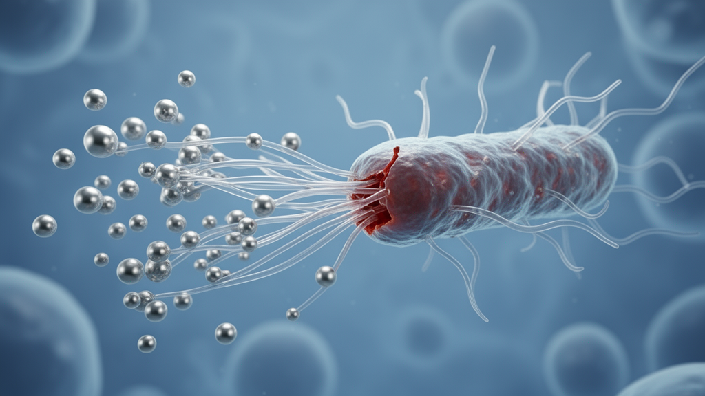
Antibiotic resistance is quietly becoming one of the defining medical crises of our time, and now a team of researchers from Italy, the United States, and Pakistan has unveiled a strikingly elegant solution that sounds like it was plucked straight from a garden. In a study published in Frontiers in Natural Products, scientists transformed discarded Camellia japonica flower petals into a powerful antibacterial weapon by using their natural polyphenols to synthesize silver nanoparticles, then pairing those particles with engineered cationic polymers to create hybrid nanoassemblies that fight multidrug-resistant bacteria more effectively than either component alone.
The research, led by Humaira Khan and colleagues at the University of Campania Luigi Vanvitelli and the University of Massachusetts Amherst, tackles a persistent challenge in modern medicine: Gram-negative bacteria like Acinetobacter baumannii and Escherichia coli, which the World Health Organization has flagged as priority pathogens, are increasingly shrugging off conventional antibiotics. These bacteria possess a formidable outer membrane that blocks many drugs from penetrating, and their rising resistance has left clinicians scrambling for alternatives. The new study offers a materials-science answer, combining two complementary antimicrobial mechanisms within a single nanoscale package.
What makes the approach so compelling is its sustainability. Rather than relying on harsh chemical reducing agents like sodium borohydride, which can compromise biocompatibility and burden the environment, the researchers harnessed the natural chemistry of Camellia japonica flowers. Using ultrasound-assisted extraction with formic acid in ethanol, they recovered a polyphenol-rich extract from freeze-dried petals, achieving a 75 percent crude yield. Subsequent purification on Amberlite XAD-4 resin concentrated the polyphenolic fraction to 94 milligrams per gram of dried flowers.
Comprehensive chemical profiling using UHPLC-HRMS/MS and HPLC-DAD revealed that the extract is dominated by anthocyanins, particularly cyanidin derivatives, alongside flavan-3-ols like catechin and epicatechin, oligomeric procyanidins, and other flavonoids. Cyanidin 3-O-glucoside and its p-coumaroyl derivative emerged as the main pigments. These compounds are not merely passengers; they are the active workforce. When the researchers mixed the extract with silver nitrate solution at pH 10 and 60 degrees Celsius, the phenolic hydroxyl groups donated electrons to silver ions, reducing them to metallic silver atoms that nucleated into nanoparticles. Simultaneously, residual polyphenols adsorbed onto the particle surfaces, acting as natural capping agents that prevented aggregation.
The spectroscopic fingerprints told the story clearly. The UV-Vis spectrum of the resulting colloidal suspension displayed a sharp, symmetric surface plasmon resonance band centered at 400 nanometers, characteristic of small, nearly spherical silver nanoparticles. Meanwhile, the intense 280-nanometer polyphenol band diminished and the 528-nanometer anthocyanin band vanished entirely, confirming that the most abundant pigments had participated in the reduction and capping process. ATR-FTIR spectroscopy reinforced this picture, showing shifts in hydroxyl, aromatic, and carbonyl bands after nanoparticle formation, while citrate-stabilized chemically synthesized AgNPs prepared for comparison exhibited a fundamentally different surface chemistry dominated by carboxylate vibrations.
Transmission electron microscopy revealed predominantly spherical particles with diameters of 20 to 30 nanometers and a narrow size distribution, consistent with the sharp SPR band and indicating effective stabilization by the plant-derived coating. Dynamic light scattering measured a hydrodynamic diameter of 66 nanometers for the AgNPs, with a negative zeta potential of -21.5 millivolts, typical of phytochemical-capped silver colloids. The green-synthesized particles also maintained measurable radical-scavenging activity at low concentrations in ABTS assays, where chemically synthesized counterparts showed markedly reduced effectiveness, suggesting the polyphenol coating confers additional functional benefits beyond stabilization.
On the biological front, the polyphenol-capped AgNPs inhibited the growth of multidrug-resistant A. baumannii and E. coli at minimum inhibitory concentrations of 10 micrograms per milliliter, while the cationic PONI-C11-TMA polymeric nanoparticles, which mimic host-defense peptides and kill bacteria by disrupting membranes, achieved inhibition at 2.5 micrograms per milliliter. Crucially, cytotoxicity testing on NIH-3T3 fibroblast cells showed viability remained above 90 percent even at 70 micrograms per milliliter, far exceeding the effective antimicrobial doses. The natural polyphenol capping appears to moderate silver ion release, minimizing collateral damage to mammalian cells while preserving bacterial selectivity.
The real breakthrough came when the researchers combined the two nanomaterials. Positively charged PONI-C11-TMA nanoparticles, with a zeta potential of +21.3 millivolts, spontaneously assembled with the negatively charged AgNPs through electrostatic interactions between quaternary ammonium groups and anionic phytochemical moieties. The resulting hybrid PNP-Ag nanoassemblies exhibited an increased hydrodynamic diameter of 91.1 nanometers and a moderated positive surface charge of +5.1 millivolts, confirming successful incorporation of AgNPs within the polymeric matrix without macroscopic aggregation.
Checkerboard titration against the resistant strains revealed that the hybrid nanoassemblies reduced effective antimicrobial concentrations by 2 to 4 fold against A. baumannii and 4 to 8 fold against E. coli, with a fractional inhibitory concentration index of 0.75, indicating an additive interaction. This means the two mechanisms, electrostatic membrane disruption by the cationic polymer and reactive oxygen species generation by the silver core, work in concert rather than redundantly. The species-specific differences in MIC reduction likely reflect variations in membrane composition or stress response pathways between the two pathogens.
Beyond the immediate antibacterial results, the study carries broader implications. Post-blooming floral waste, typically discarded as horticultural byproduct, represents an underutilized reservoir of bioactive polyphenols. By valorizing this waste stream into functional nanomaterials, the researchers align their work with circular economy principles and the United Nations Sustainable Development Goals. The modular nature of the platform, where different plant extracts could be swapped in and various cationic polymers paired out, suggests a flexible design space for future antimicrobial nanomedicines. While the authors acknowledge that further studies using human-derived cell models, extended exposure periods, and direct evaluation of the hybrid assemblies themselves will be needed to fully characterize long-term biocompatibility, the current findings establish a promising foundation. As multidrug-resistant infections continue to outpace conventional antibiotic development, hybrid nanoassemblies built from flower petals and engineered polymers offer a glimpse of how sustainable chemistry and precision materials design might converge to address one of medicine’s most stubborn challenges.
Subject of Research: Green-synthesized silver nanoparticles from Camellia japonica flowers combined with cationic polymers form additive antibacterial hybrid nanoassemblies
Article Title: Phytochemical-capped silver nanoparticles and cationic polymeric nanoparticles form antibacterial hybrid nanoassemblies
Article References: Khan, H., Abdelaziz, M., Nasim, N., Park, J., Piccolella, S., Rotello, V. M., & Pacifico, S. (2026). Phytochemical-capped silver nanoparticles and cationic polymeric nanoparticles form antibacterial hybrid nanoassemblies. Frontiers in Natural Products, 5, Article 1955630. https://doi.org/10.3389/fntpr.2026.1955630
Image Credits: AI Generated
DOI: 10.3389/fntpr.2026.1955630
Keywords: antibacterial, silver nanoparticles, Camellia japonica, hybrid nanoassemblies, green synthesis, antimicrobial resistance, polyphenols, cationic polymers, Acinetobacter baumannii, Escherichia coli, nanotechnology, sustainable chemistry
Cite Scienmag News
Kristina Jarvis. (October 16, 2026). Flower-Powered Silver Nanoparticles Team Up With Membrane-Attacking Polymers to Fight Superbugs. Scienmag. https://scienmag.com/flower-powered-silver-nanoparticles-team-up-with-membrane-attacking-polymers-to-fight-superbugs/
Kristina Jarvis. "Flower-Powered Silver Nanoparticles Team Up With Membrane-Attacking Polymers to Fight Superbugs." Scienmag, 16 October 2026, https://scienmag.com/flower-powered-silver-nanoparticles-team-up-with-membrane-attacking-polymers-to-fight-superbugs/. Accessed 16 October 2026.
Kristina Jarvis. "Flower-Powered Silver Nanoparticles Team Up With Membrane-Attacking Polymers to Fight Superbugs." Scienmag. October 16, 2026. https://scienmag.com/flower-powered-silver-nanoparticles-team-up-with-membrane-attacking-polymers-to-fight-superbugs/


