


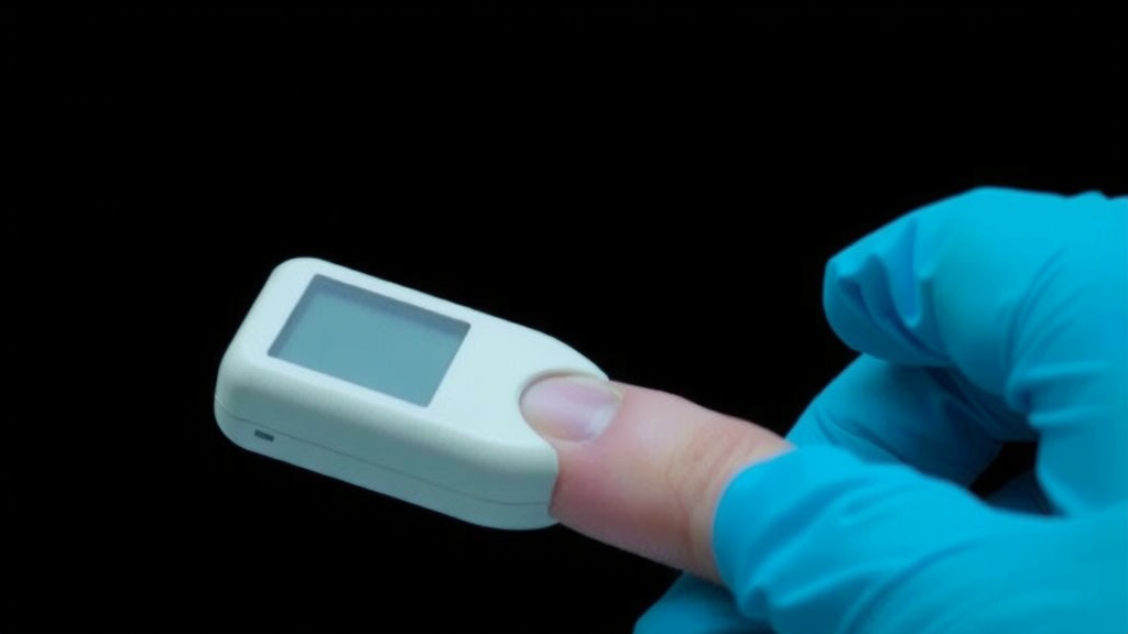
Traumatic brain injury affects roughly 69 million people each year worldwide and costs health systems more than $400 billion annually, yet diagnosing and monitoring it still depends on infrastructure that many clinics and field settings simply do not have. Current approaches rely on CT imaging, intracranial pressure monitoring, or blood-based biomarker tests that demand rapid processing, refrigeration, and in many cases venipuncture by trained personnel. Long-term biobanking pushes the burden even further, requiring serum or plasma to be frozen at minus 80 degrees Celsius to prevent degradation. A research team led by Firas Kobeissy and Kevin K. Wang, publishing in the Journal of Advanced Research, now reports the evaluation of a novel device that could loosen these constraints dramatically: a Dried Plasma Biosampling Lateral Flow (DPB-LF) device that captures plasma from a simple finger-prick of blood, dries it on a membrane, and preserves key brain-injury proteins for days without a freezer.
The device borrows its core architecture from the lateral flow assay, the same simple, fast technology behind home pregnancy tests and decades of glucose monitoring. Built from components supplied by lateral flow specialist DCN Dx, the DPB-LF cassette sandwiches a 9 mm plastic-backed nitrocellulose membrane between two functional zones. On one side, a Cytosep HV Plus wick transports fluid by capillary action; on the other, a Vivid Cell/Plasma Separation membrane filters blood cells, debris, and particles out of an applied capillary blood sample, allowing only the plasma fraction to migrate onto and bind within the central nitrocellulose collection zone. A window in the closed casing lets users visually confirm that plasma has been retained, appearing as a light yellow to red patch, while dark red dried blood cells remain trapped at the sample well by the cell filter.
Importantly, the researchers deliberately avoided imposing a tight molecular weight cutoff on the device. Although the nitrocellulose zone typically retains molecules below roughly 50 kilodaltons, the team notes that several critical TBI biomarkers circulate in oligomeric forms exceeding 100 kilodaltons, including glial fibrillary acidic protein (GFAP) and Tau, as well as alphaII-spectrin breakdown products such as SBDP150 and SBDP120. A restrictive cutoff would therefore risk losing exactly the proteins clinicians most want to measure. Once assembled, the composite membrane structure is cut into 5 mm-wide strips and housed in MICA 200 cassettes, producing a self-contained unit that a patient, caregiver, or field medic can load with a fingerstick sample in seconds.
To test whether dried plasma could faithfully preserve the protein content of liquid samples, the team first applied fresh capillary blood from healthy volunteers to the devices, air-dried them, and then rehydrated the membranes. SDS-PAGE analysis with Coomassie Blue staining revealed distinct, consistent protein banding patterns spanning roughly 200 kilodaltons down to 10 kilodaltons, closely matching patterns obtained from conventional wet venous plasma. The main variation between individuals appeared in the hemoglobin band at 16 kilodaltons, reflecting differences in hemolysis tendency during fingerstick collection rather than device failure. Quantitative testing using ten archived control and ten TBI plasma samples showed average total protein recovery of 59.91 percent for controls and 57.02 percent for TBI samples after drying and rehydration, figures the researchers consider adequate for downstream immunoassay work.
The central stability experiment asked how well four established TBI biomarkers, GFAP, neurofilament light (NfL), Tau, and ubiquitin carboxy-terminal hydrolase-L1 (UCH-L1), survive drying and storage. Pooled plasma from 20 TBI patients and 20 healthy controls was loaded onto devices, sealed in aluminum bags with desiccant, and stored at 4 degrees Celsius, room temperature of 22 to 25 degrees Celsius, or a thermal stress condition of 40 degrees Celsius for 1, 7, 14, or 21 days. After rehydration in CHAPS buffer, biomarkers were quantified using the ultrasensitive Quanterix single-molecule array platform. The verdict was clear: refrigeration at 4 degrees Celsius preserved TBI biomarker levels essentially unchanged for the full 21 days, with GFAP holding steady between 1,500 and 1,600 pg/mL and NfL and Tau remaining flat.
Room temperature proved acceptable for shorter periods, with no appreciable loss of biomarker signal through seven days of storage, a window the authors identify as the most realistic operational scenario for field deployment before samples reach a laboratory. By day 21 at room temperature, however, TBI sample levels had declined substantially, with GFAP falling from over 2,000 pg/mL to 775 pg/mL. The 40 degree Celsius stress test, designed to model extreme austere environments, produced steep losses by day 21: GFAP dropped roughly 69 percent, NfL about 78 percent, and Tau around 86 percent, although signals were never completely abolished, suggesting that improved desiccants, membrane chemistries, and barrier materials could extend thermal robustness in future designs.
Validation in real patient cohorts followed. In the TRACK-GERI pilot study of older adults with TBI, 16 geriatric patients aged 65 to 92 yielded paired wet and dried plasma samples. Average total protein recovery reached 91.6 percent, and biomarker recovery was striking: median recovery was 93 percent for GFAP, 104 percent for NfL, 173 percent for Tau, and 381 percent for UCH-L1. Recovery values exceeding 100 percent do not mean the dried samples contain more protein; the authors attribute the elevated apparent signal to an antigen-revealing effect analogous to antigen retrieval in immunohistochemistry, in which drying and migration across the membrane remove interfering plasma constituents or soluble binding partners and unmask epitopes for better antibody access. Crucially, wet and dried measurements remained strongly correlated across paired samples, with R-squared values of 0.995 for GFAP, 0.991 for NfL, 0.980 for Tau, and a weaker but still meaningful 0.829 for UCH-L1. Notably, four wet plasma samples failed to yield usable assay data, likely due to lipid interference, while their dried counterparts produced results, hinting that the drying step may actually rescue problematic samples.
A second, independent cohort of 44 TBI subjects from Baylor College of Medicine, spanning all severities from Glasgow Coma Scale 3 to 15 and including 28 CT-positive and 16 CT-negative patients, extended the findings. All four biomarkers were significantly elevated in CT-positive versus CT-negative patients in both dried and wet formats, and receiver operating characteristic analysis showed moderate-to-good diagnostic discrimination for rehydrated dried plasma, with area under the curve values of 0.756 for GFAP, 0.734 for NfL, 0.738 for Tau, and 0.703 for UCH-L1. Wet plasma performed slightly better, with GFAP reaching an AUC of 0.868, but paired bootstrap comparisons found the wet-versus-dried differences were not statistically significant for any biomarker. Biomarker concentrations also tracked injury severity across GCS categories in both formats, rising stepwise from mild to severe injury, with the sole exception of NfL measured from dried plasma, which narrowly missed significance.
The clinical implications reach well beyond the laboratory. Because the device requires only a fingerstick, the same minimally invasive technique diabetes patients have used at home for years, it sidesteps venipuncture entirely, a major advantage for elderly patients with fragile veins, patients in hypovolemic shock with compromised vascular access, or people with collapsed veins. Samples dried on the device tolerate up to a week at room temperature and can be shipped without cold chains, centrifugation, or immediate processing, enabling decentralized collection at homes, ambulances, or austere field sites with delayed centralized analysis. The authors envision event-triggered monitoring after falls or suspected head trauma, retrospective diagnosis in older adults discharged from emergency departments, and longitudinal tracking of recovery, particularly valuable given that half of the geriatric cohort had pre-existing mild cognitive impairment or dementia that complicates clinical assessment.
The team is careful to frame the work as analytical feasibility rather than a finished diagnostic. Formal regulatory evaluation, likely through an FDA clearance pathway for blood collection and pre-analytical handling devices, lies ahead, as does validation in larger and more demographically diverse cohorts. UCH-L1 remains the weakest link, showing the poorest wet-to-dried correlation and possibly interacting with hemoglobin, a question the authors flag for further study. Yet the core demonstration stands: a credit-card-sized lateral flow cassette can separate plasma from a drop of fingerstick blood, dry it, and preserve the molecular signatures of brain injury with enough fidelity to match ultrasensitive laboratory assays. If future validation confirms these results, the humble technology behind the pregnancy test could bring sophisticated brain-injury biomarker testing to ambulances, sports sidelines, rural clinics, and patients’ own homes, transforming a diagnostic workflow that has long demanded freezers, phlebotomists, and haste into one that fits in a shirt pocket.
Subject of Research: A dried plasma lateral flow biosampling device for stabilizing traumatic brain injury protein biomarkers
Article Title: Evaluation of a novel high-capacity dry plasma biosampling-lateral flow device for the testing of traumatic brain injury protein biomarkers
Article References: Kobeissy, F., Elbayoumi, E., Arja, R. D., Cai, G., Boukholda, K., Apiliogullari, S., Nelson, M. D., Yadikar, H., Alawieh, A. M., Grossberg, J. A., Mechref, Y., Wagner, A. K., Rubenstein, R., Robertson, C., Yamal, J.-M., Tsetsou, S., Gardner, R. C., & Wang, K. K. (2026). Evaluation of a novel high-capacity dry plasma biosampling-lateral flow device for the testing of traumatic brain injury protein biomarkers. Journal of Advanced Research. https://doi.org/10.1016/j.jare.2026.09.003
Image Credits: AI Generated
DOI: 10.1016/j.jare.2026.09.003
Keywords: traumatic brain injury, biomarkers, lateral flow assay, dried plasma, GFAP, UCH-L1, neurofilament light, Tau, point-of-care diagnostics, biosampling, protein stability, fingerstick blood
Cite Scienmag News
Cassandra Pierce. (September 24, 2026). Finger-Prick Device Keeps Brain Injury Biomarkers Stable Without Freezers. Scienmag. https://scienmag.com/finger-prick-device-keeps-brain-injury-biomarkers-stable-without-freezers/
Cassandra Pierce. "Finger-Prick Device Keeps Brain Injury Biomarkers Stable Without Freezers." Scienmag, 24 September 2026, https://scienmag.com/finger-prick-device-keeps-brain-injury-biomarkers-stable-without-freezers/. Accessed 4 October 2026.
Cassandra Pierce. "Finger-Prick Device Keeps Brain Injury Biomarkers Stable Without Freezers." Scienmag. September 24, 2026. https://scienmag.com/finger-prick-device-keeps-brain-injury-biomarkers-stable-without-freezers/


