


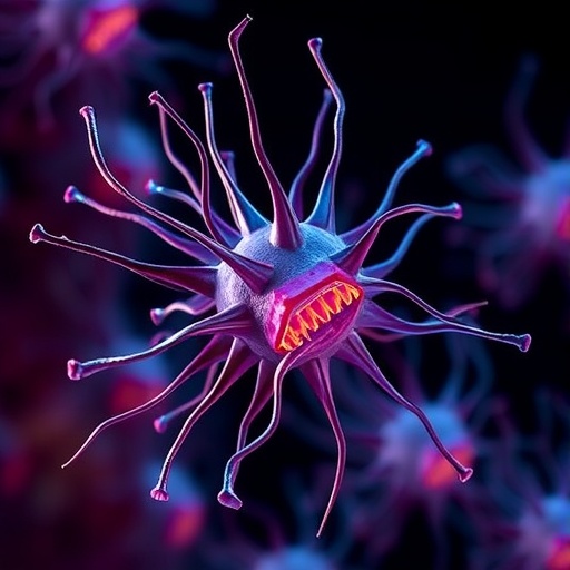
In the vast, unseen world beneath the ocean’s surface, planktonic organisms represent one of the most fundamental engines of life on Earth. Responsible for generating a significant portion of the planet’s oxygen and anchoring the marine food web, plankton encompass an enormous variety of species, from tiny photosynthesizing algae to diverse protists. Among these, protists—the intricately complex, single-celled eukaryotic microorganisms—hold particular evolutionary importance. However, despite decades of genomic explorations, a critical gap remained in our ability to visualize their cellular structures, mainly due to technical limitations in microscopy methods for intact imaging. This gap is now being revolutionized by an innovative microscopy technique adapted and refined over the last few years, allowing researchers to peer into the ultrastructure of protists with unprecedented clarity.
The breakthrough came from a remarkable collaboration between European Molecular Biology Laboratory (EMBL) Group Leader Gautam Dey, Omaya Dudin—then at EPFL—and scientists Paul Guichard and Virginie Hamel at the University of Geneva. They leveraged the pioneering technology of expansion microscopy, originally developed at MIT, and further optimized it into Ultrastructure Expansion Microscopy (U-ExM). This technique effectively renders otherwise impermeable cell walls porous, enabling fluorescent labeling and imaging of intracellular components that were once inaccessible. When Dudin first applied this to Ichthyosporea, a marine protist linked closely to animals and fungi, they successfully visualized its internal architecture, breaking a longstanding barrier in protistology.
The ambition of this team extended far beyond single species studies. They forged an international collaboration that recently culminated in exploring the cellular organization of over 200 eukaryotic plankton species—a monumental endeavor documented in the journal Cell. This project, part of the Traversing European Coastlines (TREC) expedition and dubbed PlanExM, demonstrates a stunning panorama of ultrastructural diversity across marine microbes. It’s arguably the first comprehensive atlas to detail the microscopic landscapes that underpin life at the cellular level in these enigmatic organisms, opening up new vistas for evolutionary biology and cell science.
One of the key sites fueling this research was the Station Biologique in Roscoff, France, a premier marine research facility housing one of Europe’s most exhaustive culture collections of marine microorganisms. Upon requesting samples for testing expansion microscopy, the researchers were astounded to find access granted to over 200 unique species, far surpassing expectations. According to co-first author Felix Mikus, this “treasure trove” allowed them to fix and prepare samples intensively over several continuous days, marking a pivotal moment in accessing previously hidden cellular intricacies.
Expansion microscopy itself is a technological marvel that circumvents the classical diffraction limits of light microscopy. By embedding biological specimens in a swellable hydrogel, and inducing their physical expansion, spatial resolution is boosted dramatically. This expansion—ranging typically from fourfold to sixteenfold linear increase—preserves the relative positions of biomolecules, allowing structural details at nanometer scales to be resolved with standard light microscopy setups. This technique sidesteps the need for costly super-resolution microscopes and complex optical setups, democratizing access to ultrastructural details across biological disciplines.
Harnessing this technique, researchers homed in on the cytoskeleton, the intricate filamentous network that shapes the interior scaffolding of eukaryotic cells. In particular, they examined microtubules—rigid, hollow tubes vital for cell shape, division, and motility—and centrins, specialized proteins instrumental in organizing microtubule architecture within microtubule-organizing centers. The scope of their investigation, spanning hundreds of species from diverse clades, enabled mapping the variations in cytoskeletal organization, uncovering both conserved and distinctive traits across the eukaryotic domain.
Hiral Shah, an EMBL Postdoctoral Fellow, emphasized how these patterns of tubulin and centrin organization across various groups allow not only a snapshot of structural diversity but also evolutionary predictions. For example, dinoflagellates—a speciose and ecologically critical group in marine environments—displayed distinctive tubulin and centrin structures linked to cell cortical regions and flagella, offering clues to their evolutionary adaptations. Such information was previously unattainable with traditional microscopy, highlighting the transformative potential of combining U-ExM with large-scale biodiversity studies.
Armando Rubio Ramos, also a co-first author and researcher at the University of Geneva, remarked on the paradigm shift enabled by this ultrastructural expansion approach. Integrating U-ExM with high-throughput imaging platforms and computational comparative analyses bridges the gap between molecular sequence data and physical cellular organization. This fusion creates a new dimension for interpreting how subcellular architectures have evolved and diversified, laying a foundation for exploring cellular morphology in an evolutionary context across microbial eukaryotes.
Beyond fundamental biological insights, the research demonstrates the practicality of expansion microscopy for direct analysis of complex environmental samples. The technique’s adaptability to natural plankton collected from marine ecosystems sidesteps challenges posed by cell wall impermeability, heterogeneous cell populations, and operational complexity. This opens the door to broad application prospects, such as monitoring ecological dynamics, assessing biodiversity shifts, and linking cellular physiology with environmental variables in real-time.
The success of this interdisciplinary endeavor has attracted significant support, underscored by securing a CHF 2 million grant from the prestigious Moore Foundation, with contributions from collaborators including Thomas Richards from Oxford University. This funding paves the way for deeper exploration into selected protist species to address fundamental questions about cell division mechanisms, the emergence of multicellularity, and phenotypic diversity underpinning evolutionary transitions. Such studies hold promise for integrating microscopy-derived phenotypes with multiomics datasets, revolutionizing our understanding of life’s cellular foundations at scale.
Gautam Dey envisions expansion microscopy becoming the first high-resolution imaging technology capable of matching the scale and depth of contemporary biodiversity genomics projects. This synergy could facilitate associating genetic and molecular data with detailed cellular physiology across the vast tree of life. The team’s extraordinary journey, from adapting a novel imaging method to developing a planetary atlas of microbial cytoskeletal diversity, heralds an exciting era where microscopic form and function converge to reveal life’s hidden architectures in stunning detail.
With this powerful approach now established, the research community stands at the threshold of unprecedented discovery opportunities. The fusion of expansion microscopy, high-throughput imaging, and evolutionary biology promises to unravel the deepest secrets of microbial eukaryotes, elucidating how cellular complexity evolved, diversified, and impacts ecosystems worldwide. As the project moves forward, it symbolizes a landmark shift in the visualization and understanding of the invisible majority of life beneath the waves.
- TREC Expedition
- Moore Foundation Grant Announcement
References: - DOI: 10.1016/j.cell.2025.09.027
Image Credits: Felix Mikus/EMBL
Keywords: Molecular biology, Cell biology
Cite Scienmag News
Violet Maxwell. (October 31, 2025). Expansion Microscopy Unveils the Hidden World of Plankton. Scienmag. https://scienmag.com/expansion-microscopy-unveils-the-hidden-world-of-plankton/
Violet Maxwell. "Expansion Microscopy Unveils the Hidden World of Plankton." Scienmag, 31 October 2025, https://scienmag.com/expansion-microscopy-unveils-the-hidden-world-of-plankton/. Accessed 3 September 2026.
Violet Maxwell. "Expansion Microscopy Unveils the Hidden World of Plankton." Scienmag. October 31, 2025. https://scienmag.com/expansion-microscopy-unveils-the-hidden-world-of-plankton/


