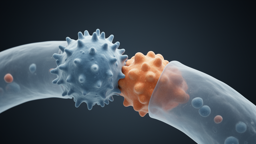
Scientists at Albert Einstein College of Medicine, working with collaborators at Institut Pasteur and LSU Health Shreveport, have published a study in Nature showing how the Andes hantavirus recognizes its target on a human cell. The work arrives against a sobering backdrop: earlier this year, an outbreak of Andes hantavirus on a cruise ship killed three passengers and sickened 13, underscoring the absence of approved vaccines or treatments for the virus. The new findings, the researchers say, could inform the development of effective therapies against Andes virus and other hantaviruses.
Hantaviruses are a large family of viruses carried mainly by rodents, and they can cause serious and sometimes fatal diseases in people, including hantavirus pulmonary syndrome, a severe respiratory illness that can rapidly lead to breathing failure. Human infections occur mainly when people inhale airborne virus particles from the urine, droppings, or saliva of infected rodents. Of the more than 20 hantaviruses known to sicken humans, Andes virus is the only one known to spread from person to person, typically through close and prolonged contact. This year’s cruise ship outbreak provided a dramatic example of that capability.
The research team had previously established that infection depends on the spike protein on the Andes virus surface latching onto a receptor called PCDH1 on the surface of human cells. That receptor essentially serves as a doorway for the virus to enter the cell and then multiply inside it. What remained unanswered was a precise mechanistic question: how does the spike protein actually latch onto PCDH1? Eight years ago the group discovered that Andes virus uses this particular surface protein to infect human cells, but the exact recognition mechanism eluded them.
“Eight years ago, we discovered that Andes virus infects human cells by using a particular protein on the cell’s surface, called a receptor, but we couldn’t figure out exactly how the virus recognized it,” said co-corresponding author Kartik Chandran, Ph.D., professor of microbiology and immunology at Einstein. “Our latest work solves this puzzle: We reveal a new way in which a virus can capture its receptor during infection and also suggest how we can fight back with therapies targeted to exploit the resulting viral vulnerability.”
The answer lies in an unusual architecture. The spike protein of SARS-CoV-2, the virus that causes COVID-19, is built from three identical protein units arranged in a triangle, each carrying a separate receptor-binding domain that latches onto its receptor, ACE2. The Andes virus spike, by contrast, is an arrangement of four identical protein units locked together into a square. Unlike the SARS-CoV-2 and ACE2 pairing, a single protein unit from the Andes virus cannot bind to its receptor on its own; instead, the entire square-shaped spike is required to capture PCDH1.
To see what happens at the very beginning of infection, when the Andes virus first encounters a human cell, the researchers used cryo-electron microscopy to reveal the structure in sharp detail. That step was accomplished only after the team developed innovative new methods, including an artificial intelligence-designed scaffold, that enabled the square-shaped Andes virus spike to hold together in a test tube. “The best way to prove a rare bird exists is to take a picture of it,” said Pablo Guardado-Calvo, Ph.D., deputy director of the Virology Department and head of the Structural Biology of Infectious Diseases unit at Institut Pasteur. “When we mixed this engineered Andes virus spike with PCDH1 and looked at it under the microscope, we knew we had something special.”
The structures showed that a single molecule of PCDH1 embeds itself into the heart of the spike by binding into a deep groove, or pocket, at its center. The researchers also resolved molecular specifics of the interaction that will be important for designing treatments intended to block it. “The depth of this pocket also helps explain how the virus limits antibody access,” said Rohit Jangra, Ph.D., assistant professor of microbiology and immunology at LSU Health Shreveport. “What’s striking is how the same pocket can accommodate the receptor while restricting antibody access. PCDH1’s long, slender shape lets it reach deep inside, but most antibodies are too bulky to follow.”
The team then examined several antibodies isolated from human hantavirus patients that had previously been shown to block the Andes virus-PCDH1 binding interaction. They found that some of these antibodies could indeed bind close enough to the entrance of the receptor-binding pocket to prevent PCDH1 from entering it, but they could not fit as deeply into the pocket as PCDH1 does. According to Chandran, this suggests a clever way in which Andes virus can escape receptor-blocking antibodies: the virus mutates so that antibodies cannot destroy it, yet those changes do not affect its ability to bind to the receptor, a kind of molecular “friend-or-foe” identification system.
That same antibody-escape strategy, however, creates a vulnerability the team intends to exploit. Although human antibodies are limited to a certain architecture, scientists can create “long and skinny” molecules, suggesting an approach that could block the PCDH1 pocket and offer a blueprint for the development of new antiviral medicines that the researchers describe as urgently needed for Andes virus and its close relative in the United States and Canada, Sin Nombre virus. “This work creates a unified picture of the hantavirus-receptor interaction that could help us conceive and discover new protective strategies that work broadly against these high-consequence pathogens,” said Dr. Guardado-Calvo.
The paper, titled “Structural basis for protocadherin-1 recognition by New World hantaviruses,” lists Megan M. Slough, Ph.D., of Albert Einstein College of Medicine as first author, with co-authors including Leandro Battini, Ph.D., of Institut Pasteur and Ramandeep Kaur, Ph.D., of LSU Health Shreveport, along with additional contributors from The Henry M. Jackson Foundation, The University of Texas at Austin, University College London, Ichor Biologics LLC, Norgen Biotek, Universidad San Sebastián, the U.S. Army Medical Research Institute of Infectious Diseases, and Fundacion Ciencia & Vida. The work was funded by the National Institutes of Health, through grant U19AI181977 (“PROVIDENT”), and by the European Union (EVH, GAP-101202831). As a structural study, it defines the molecular mechanics of receptor recognition and points to candidate therapeutic strategies, which would still need to be developed and tested before any clinical benefit could be established.
Subject of Research: Structural basis of Andes hantavirus recognition of the PCDH1 receptor
Article Title: Scientists reveal how deadly Andes hantavirus attaches to human cells
Article References: Scientists reveal how deadly Andes hantavirus attaches to human cells. Original publication (Bibliographic details unavailable.)
Image Credits: AI Generated
DOI: Not provided
Keywords: Andes virus, hantavirus, PCDH1, cryo-electron microscopy, structural biology, spike protein, antibody escape, Sin Nombre virus, Institut Pasteur, Albert Einstein College of Medicine, Nature, antiviral targets
Cite Scienmag News
Drew Townsend. (October 28, 2026). Cryo-EM reveals how Andes hantavirus grips its human cell receptor. Scienmag. https://scienmag.com/cryo-em-reveals-how-andes-hantavirus-grips-its-human-cell-receptor/
Drew Townsend. "Cryo-EM reveals how Andes hantavirus grips its human cell receptor." Scienmag, 28 October 2026, https://scienmag.com/cryo-em-reveals-how-andes-hantavirus-grips-its-human-cell-receptor/. Accessed 28 October 2026.
Drew Townsend. "Cryo-EM reveals how Andes hantavirus grips its human cell receptor." Scienmag. October 28, 2026. https://scienmag.com/cryo-em-reveals-how-andes-hantavirus-grips-its-human-cell-receptor/