


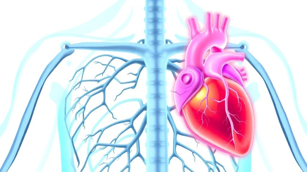
Every heartbeat depends on a hidden piece of fluid engineering. When blood rushes through the mitral valve into the left ventricle, it ideally curls into a coherent, ring-shaped vortex that steers the incoming jet toward the outflow tract and keeps energy losses to a minimum. A new computational study published in the Annals of Biomedical Engineering suggests that when the mitral valve narrows, this elegant vortex collapses, and that the way it collapses depends strikingly on what caused the narrowing in the first place. The work, led by Marshall Davey and Boyce Griffith of the University of North Carolina at Chapel Hill together with Mohammad Saber Hashemi, Gregg Pressman, and Arash Kheradvar, is among the first to compare the two dominant forms of mitral stenosis using fully three-dimensional, patient-specific fluid-structure interaction simulations.
Mitral stenosis accounts for roughly twelve percent of patients with disease confined to a single heart valve, and it comes in two very different flavors. Rheumatic mitral stenosis, long the dominant form worldwide, arises from repeated group A streptococcal infections. Molecular mimicry between bacterial antigens and valve proteins triggers an autoimmune attack that fibroses the leaflets and fuses their commissures, sculpting the valve into a funnel whose tightest point sits at the leaflet tips. Mitral annular calcification, by contrast, is an inflammatory disease of aging in which calcium deposits build up in the fibrous ring anchoring the valve, sometimes extending onto the leaflet bases and forming a rigid calcific tunnel. In this condition the narrowest flow channel lies near the annulus rather than at the tips, and the entire valve apparatus takes on a shrunken appearance. In a large United States echocardiographic cohort, annular calcification was found in twenty-three percent of imaged patients, and stenosis was present in 6.6 percent of those with calcification compared with just 0.5 percent of those without. Prognosis is sobering: five-year survival in mitral stenosis associated with annular calcification has been reported below fifty percent.
Clinical imaging struggles to distinguish what these two diseases actually do to the blood. Doppler echocardiography measures velocities only along the ultrasound beam, so it cannot reconstruct the full three-dimensional velocity field inside the ventricle, and even four-dimensional flow magnetic resonance imaging lacks the resolution to capture the fine shear layers where viscous energy is dissipated. Earlier laboratory work using planar particle image velocimetry had hinted that calcific stenosis dissipates more energy than the rheumatic form, but a single imaging plane can miss out-of-plane velocities entirely. Because ventricular flow is inherently three-dimensional, the team turned to high-fidelity computation to see what planar measurements might be hiding.
The researchers built their virtual laboratory around real patients. Three mitral valve geometries, one healthy, one rheumatic, and one calcific, were digitally extracted from four-dimensional echocardiography at the moment of systolic closure, thickened to one millimeter, and embedded in a left ventricle model derived from a silicone phantom used in prior bench experiments. The calcific case included an additional rigid ring representing the annular calcium. Using an immersed finite element-finite difference fluid-structure interaction scheme with adaptive mesh refinement, the team simulated eight consecutive cardiac cycles at seventy beats per minute across cardiac outputs of 1.5, 3.0, 3.5, and 5.0 liters per minute, discarding the first three cycles so that statistics were drawn from a periodic steady state. Blood was modeled as an incompressible Newtonian fluid with a viscosity of 4.0 millipascal-seconds, and parallel simulations with water-like viscosity allowed direct comparison with the original in vitro platform.
The classic diagnostic numbers told a familiar but incomplete story. Both diseased valves produced elevated transmitral pressure gradients relative to the healthy valve, as expected. The rheumatic valve had the smallest geometric and effective orifice areas, yet the differences in mean pressure gradient and peak velocity between the two diseases flipped depending on cardiac output: peak velocity was higher in the rheumatic case at reduced flows, but the calcific valve took the lead at the physiologic 5.0 liters per minute. When the gradients were scaled by orifice area, however, the calcific valve carried the heavier effective hemodynamic burden, consistent with earlier bench findings.
The energetics told a sharper tale. Despite the rheumatic valve’s tighter orifice and higher velocities, the calcific case demanded the greatest pump stroke work, the mechanical energy the ventricle must expend to sustain each beat, and it also produced the highest three-dimensional ventricular kinetic energy and viscous energy dissipation across all equivalent flow conditions. The healthy valve sat lowest on every energetic measure. Crucially, the gap between the two diseases widened dramatically when the full three-dimensional velocity field was used instead of a single plane. At 3.5 liters per minute with blood-analog viscosity, planar measurements suggested only a twenty percent difference in total cycle dissipation between the calcific and rheumatic cases; the three-dimensional analysis put it at fifty-one percent. The discrepancy traces to large out-of-plane velocities in the calcific case that a planar laser sheet simply cannot see, a finding with direct implications for how laboratory flow studies of diseased hearts should be designed and interpreted.
Flow visualization and particle tracking revealed why the energy bills differ. In the healthy configuration, streamlines curled gracefully around the leaflets, forming the classic diastolic vortex ring that funnels blood toward the outflow tract. Both diseased valves destroyed this structure, but in characteristically different ways. The calcific jet drove straight toward the ventricular apex, scattering particles into the lower half of the chamber, while the rheumatic jet slammed into the inferolateral wall and sent particles crawling along the wall toward the apex, away from the exit. Of two thousand massless particles released upstream of the valve, seven hundred eight were ejected from the ventricle within three cycles in the healthy blood-analog simulation at 3.5 liters per minute, compared with 396 for the calcific valve and a mere 107 for the rheumatic valve, none of which had left the chamber by the third cycle in the water simulations. Retained particles in the calcific case traveled the longest total distances, a signature of inefficient recirculation, while ejected particles in the rheumatic case lingered longest inside the ventricle, indicating delayed filling and impaired mixing.
The clinical stakes are considerable. Breathlessness and exercise intolerance define valvular disease, and in rheumatic stenosis intervention is generally recommended once the orifice area falls below 1.5 square centimeters. Calcific stenosis is murkier: patients are older, carry more comorbidities, and it is often unclear whether their symptoms stem from the valve itself or from the stiff atrial and ventricular compliance that typically accompanies annular calcification. Intervention in this population is technically harder and riskier. If the valve’s geometric distortion is a primary driver of the physiological derangement, as these simulations suggest, then valve intervention may be justified even when conventional pressure gradients look unremarkable. Conversely, if chamber stiffness dominates, replacement could incur more risk than benefit. Etiology-specific hemodynamic signatures of the kind uncovered here could help clinicians make that distinction.
The authors are careful to frame the work as a proof of concept with real limitations. Each disease was represented by a single anonymized valve, so inter-patient variability remains unexplored; the leaflets were modeled as isotropic silicone, the calcification as fully rigid, and the subvalvular apparatus of chordae and papillary muscles was absent, which also limited loading conditions and caused the healthy valve model to invert at the highest flow rate. The ventricle was larger than physiological, yielding a reduced ejection fraction, and no direct volumetric validation against experiment or clinic was performed. Some quantitative discrepancies with the underlying bench study, including valve velocities and effective orifice areas, likely reflect unavailable pressure measurements used to set boundary conditions. Still, the qualitative vortex disruption and the comparative energetic rankings reproduced the in vitro observations, and extending them to three dimensions exposed differences no planar measurement could capture. The team calls for studies with larger valve cohorts, more physiological material models, and complementary validation data. If those confirm the pattern, the humble vortex, invisible to the stethoscope and only partially visible to the echocardiographer, may become a genuine target in the care of an aging population increasingly affected by calcified heart valves.
Subject of Research: Patient-specific fluid-structure interaction analysis of ventricular vortex formation and flow energetics in rheumatic versus calcific mitral stenosis
Article Title: Patient-Specific Analysis of Ventricular Vortex Formation and Flow Energetics in Rheumatic and Calcific Mitral Stenosis
Article References: Davey, M., Hashemi, M. S., Pressman, G. S., Kheradvar, A., & Griffith, B. E. (2026). Patient-Specific Analysis of Ventricular Vortex Formation and Flow Energetics in Rheumatic and Calcific Mitral Stenosis. Annals of Biomedical Engineering. https://doi.org/10.1007/s10439-026-04364-7
Image Credits: AI Generated
DOI: 10.1007/s10439-026-04364-7
Keywords: mitral stenosis, mitral annular calcification, rheumatic heart disease, fluid-structure interaction, ventricular vortex, cardiac fluid dynamics, flow energetics, viscous energy dissipation, echocardiography, left ventricle, biomedical engineering, computational modeling
Cite Scienmag News
Ophelia Keating. (October 9, 2026). Calcified Valves Drain the Heart’s Energy More Than Rheumatic Disease, Simulations Reveal. Scienmag. https://scienmag.com/calcified-valves-drain-the-hearts-energy-more-than-rheumatic-disease-simulations-reveal/
Ophelia Keating. "Calcified Valves Drain the Heart’s Energy More Than Rheumatic Disease, Simulations Reveal." Scienmag, 9 October 2026, https://scienmag.com/calcified-valves-drain-the-hearts-energy-more-than-rheumatic-disease-simulations-reveal/. Accessed 9 October 2026.
Ophelia Keating. "Calcified Valves Drain the Heart’s Energy More Than Rheumatic Disease, Simulations Reveal." Scienmag. October 9, 2026. https://scienmag.com/calcified-valves-drain-the-hearts-energy-more-than-rheumatic-disease-simulations-reveal/


