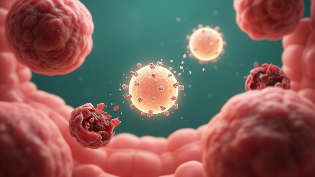
Colorectal cancer remains one of the most lethal malignancies worldwide, and the search for therapies that kill tumor cells while sparing healthy tissue has pushed researchers toward increasingly creative strategies. A team of Iranian scientists has now reported a novel twist on two converging ideas: using mesenchymal stem cells as microscopic drug factories, and using a bacterial peptide called plantaricin as the trigger that reprograms what those factories produce. Their study, published in Molecular Biology Reports, describes how tiny membrane-bound packages released by plantaricin-preconditioned stem cells can push colon cancer cells toward apoptosis, the controlled self-destruction program that healthy cells use to keep tissues in balance. The work is early-stage and entirely laboratory-based, but it offers a detailed technical blueprint for a cell-free therapeutic concept that is gaining momentum across oncology research.
The logic behind the experiment begins with mesenchymal stem cells, or MSCs, which are adult stem cells typically harvested from bone marrow. MSCs naturally shed extracellular vesicles, including microvesicles, which are spherical packets of membrane that carry proteins, lipids, and genetic cargo from the parent cell. Because these vesicles can fuse with recipient cells and deliver their contents, they have attracted intense interest as natural drug delivery vehicles. Unlike living cell therapies, vesicle-based approaches avoid the risks of uncontrolled cell division, immune rejection, and tumor initiation that have complicated stem cell transplantation. The catch is that naive MSC vesicles are not inherently toxic to cancer cells, and in some contexts MSCs have even been reported to support tumor growth. The researchers therefore set out to precondition the stem cells so that their vesicles would inherit a pro-apoptotic payload.
The preconditioning agent was plantaricin, a bacteriocin, meaning a small antimicrobial peptide produced by lactic acid bacteria such as Lactobacillus plantarum. Bacteriocins have long been valued as natural food preservatives, but a growing body of literature suggests some of them carry anticancer activity. Previous work has shown that plantaricin BM-1 reduces the viability of SW480 colorectal cancer cells by inducing caspase-dependent apoptosis, and related bacteriocins such as nisin have been shown to suppress metastatic gene expression in colon cancer cell lines. In the new study, the team exposed human bone marrow-derived MSCs to a sub-lethal dose of plantaricin, specifically 10 micrograms per milliliter, a concentration deliberately kept below the level that harms half of the cells. The idea was to stress the stem cells gently enough that they would package stress signals and apoptotic triggers into the vesicles they released, without damaging the cells themselves.
Harvesting the vesicles required differential ultracentrifugation, a technique that spins conditioned culture media at progressively higher speeds to pellet particles by size and density. The team then characterized the resulting microvesicles using three complementary methods. Flow cytometry confirmed the presence of canonical vesicle surface markers CD9, CD63, and CD81, the molecular calling cards recognized by the International Society for Extracellular Vesicles. Dynamic light scattering measured particle size distributions in suspension, and protein quantification assessed cargo concentration. The comparison between naive and plantaricin-preconditioned vesicles revealed striking physical differences. Vesicles from preconditioned cells were substantially smaller, with a mean diameter of 253 nanometers compared with 618 nanometers for naive vesicles, and they carried more protein, at 1107 versus 983 micrograms per milliliter. Smaller particles with denser cargo could, in principle, penetrate tumor tissue more efficiently and deliver a higher payload per unit volume.
The functional testing focused on HT-29 cells, a well-established human colorectal adenocarcinoma cell line, alongside HEK-293 human embryonic kidney cells as a normal-cell control. When treated with the plantaricin-enhanced vesicles, HT-29 cells showed a marked drop in viability as measured by the MTT assay, a colorimetric test in which metabolically active cells convert a yellow tetrazolium compound into a purple formazan product. The half-maximal inhibitory concentration, or IC50, for the enhanced vesicles was 39.10 micrograms per milliliter, significantly lower than the 52.43 micrograms per milliliter required for naive vesicles, a difference the authors report as highly significant. Crucially, neither vesicle type showed meaningful cytotoxicity toward the HEK-293 normal cells, with differences failing to reach statistical significance. That selectivity, if it holds up in more complex biological systems, is precisely the property that chemotherapy has struggled to achieve.
To confirm that the viability loss reflected genuine apoptosis rather than nonspecific toxicity, the researchers used Annexin V and propidium iodide staining followed by flow cytometry. Annexin V binds phosphatidylserine, a phospholipid that migrates from the inner to the outer leaflet of the cell membrane during the early stages of apoptosis, while propidium iodide enters only cells whose membranes have already broken down, marking late apoptosis or necrosis. The results showed a dose-dependent shift. Total apoptotic HT-29 cells rose from 6.37 percent in untreated controls to 8.37 percent after treatment with naive vesicles, and to 11.15 percent after treatment with the plantaricin-enhanced vesicles. The absolute percentages are modest, and the authors are careful to frame the effect as an apoptosis-associated response rather than a dramatic cell-killing event, but the trend was statistically robust and consistent across assays.
The molecular evidence strengthened the case considerably. Quantitative real-time polymerase chain reaction, or qRT-PCR, measured the expression of three genes that sit at the heart of the apoptotic machinery. P53, the famous guardian-of-the-genome transcription factor, was upregulated by as much as 3.5-fold in treated HT-29 cells. BAX, a pro-apoptotic member of the BCL-2 family that promotes mitochondrial outer membrane permeabilization, was similarly elevated. Meanwhile BCL-2, the anti-apoptotic counterweight that normally shields cells from self-destruction, was driven down to 0.4-fold of its baseline level. This shift in the BAX-to-BCL-2 ratio is a classic biochemical signature of cells being pushed toward the mitochondrial, or intrinsic, pathway of apoptosis. In effect, the enhanced vesicles appear to be reprogramming the cancer cells’ internal accounting so that the death signals outweigh the survival signals.
Why would preconditioning stem cells with a bacterial peptide change their vesicles so profoundly? The most plausible mechanism, though the study does not fully resolve it, is that sub-lethal plantaricin exposure activates stress-response pathways within the MSCs, altering the cargo they sort into budding vesicles. Plantaricin is known to disrupt bacterial membranes by forming pores, and mammalian cells exposed to it may respond by packaging membrane-active peptides, stress proteins, or regulatory RNAs into their extracellular vesicles. When those vesicles fuse with cancer cells, the altered cargo could tip the apoptotic balance. The smaller vesicle size observed after preconditioning may itself reflect altered membrane dynamics in stressed parent cells. Disentangling exactly which cargo molecules drive the effect will be an essential next step, and techniques such as proteomic and microRNA sequencing of the vesicle contents would help identify the active ingredients.
The broader context makes this line of research worth watching. Cell-free therapies built on extracellular vesicles are advancing rapidly across regenerative medicine and oncology, with engineered MSC-derived vesicles already being explored for targeted drug delivery in cardiac tissue and for cancer treatment more broadly. Related studies have shown that MSC microvesicles can induce apoptosis in acute myeloid leukemia cells, multiple myeloma cells, and ovarian cancer cells, and MSC-derived vesicles carrying microRNAs such as miR-15a and miR-34a-5p have been shown to suppress colorectal cancer through specific molecular axes. The present study adds a new tool to that toolbox: a simple, low-cost preconditioning step using a food-grade bacterial peptide to tune vesicle cargo toward tumor killing. Because bacteriocins are generally regarded as safe and are already consumed in fermented foods, the barrier to clinical translation of the preconditioning method itself is comparatively low.
Substantial caveats remain, and the authors acknowledge them implicitly by positioning the work as a foundation for further investigation. The experiments were conducted in two-dimensional cell culture, where drug penetration, immune clearance, and tumor heterogeneity do not exist. The modest increase in apoptotic cells, from roughly 6 to 11 percent, would need to be amplified considerably before any clinical relevance could be claimed, and the selectivity observed against HEK-293 cells must be validated against a broader panel of normal tissues. Animal studies and eventually organoid models will be needed to establish whether plantaricin-enhanced vesicles can reach colorectal tumors in vivo and accumulate at therapeutic concentrations. Still, the study demonstrates a technically sound proof of concept: that the therapeutic identity of stem cell vesicles can be rewritten by preconditioning the parent cells with a bacterial peptide, and that the resulting packages can selectively engage the apoptotic machinery of colon cancer cells. In a field hungry for treatments that are both effective and gentle, that is a result worth building on.
Subject of Research: Plantaricin-preconditioned stem cell microvesicles as a cell-free approach to inducing apoptosis in colorectal cancer cells
Article Title: Harnessing microvesicles from plantaricin-enhanced stem cells to trigger apoptosis in colon cancer cells
Article References: Yaghoubinezhad, A. R., Halabian, R., Nilchiani, L. S., & Najafi, A. (2026). Harnessing microvesicles from plantaricin-enhanced stem cells to trigger apoptosis in colon cancer cells. Molecular Biology Reports, 53(1), Article 1744. https://doi.org/10.1007/s11033-026-12922-4
Image Credits: AI Generated
DOI: 10.1007/s11033-026-12922-4
Keywords: microvesicles, mesenchymal stem cells, plantaricin, colorectal cancer, apoptosis, cell-free therapy, extracellular vesicles, bacteriocins, HT-29, BAX, BCL-2, drug delivery
Cite Scienmag News
Nathaniel Bowman. (October 18, 2026). Bacterial Peptide-Primed Stem Cell Vesicles Push Colon Cancer Cells Toward Suicide. Scienmag. https://scienmag.com/bacterial-peptide-primed-stem-cell-vesicles-push-colon-cancer-cells-toward-suicide/
Nathaniel Bowman. "Bacterial Peptide-Primed Stem Cell Vesicles Push Colon Cancer Cells Toward Suicide." Scienmag, 18 October 2026, https://scienmag.com/bacterial-peptide-primed-stem-cell-vesicles-push-colon-cancer-cells-toward-suicide/. Accessed 18 October 2026.
Nathaniel Bowman. "Bacterial Peptide-Primed Stem Cell Vesicles Push Colon Cancer Cells Toward Suicide." Scienmag. October 18, 2026. https://scienmag.com/bacterial-peptide-primed-stem-cell-vesicles-push-colon-cancer-cells-toward-suicide/