


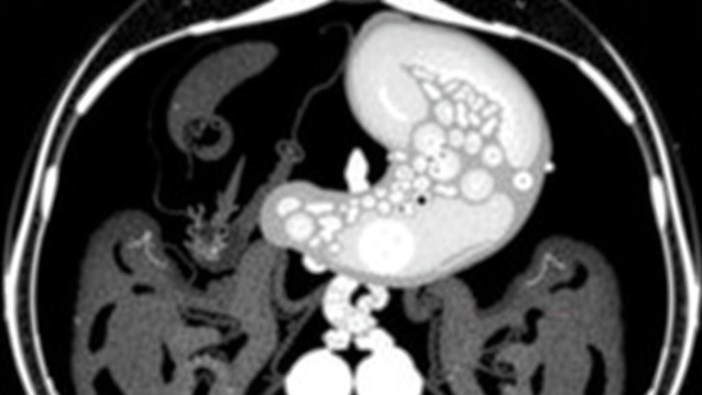
Every year, millions of people with stomach cancer face a diagnostic dilemma that goes far beyond simply locating their tumor. Once gastric cancer is confirmed, oncologists need to know the molecular character of the disease before they can choose the best treatment, and one of the most important molecular markers is a phenomenon called microsatellite instability, or MSI. Until now, determining that status has required a tissue sample taken during endoscopy or surgery, followed by laboratory testing that takes time and depends on the quality and location of the biopsy. A new multicenter study from Shanghai suggests that a routine CT scan, analyzed by machine learning, may be able to reveal this hidden molecular signature before a single cell is sampled.
The research, published in BMC Cancer by a team led by Yuqi Meng and Gang Ren of Xinhua Hospital at Shanghai Jiaotong University School of Medicine, together with Zhimeng Cui of Huadong Hospital at Fudan University, set out to answer a deceptively simple question: can the texture of a tumor as it appears on a contrast-enhanced CT image tell us whether the cancer is microsatellite instability-high? The answer, based on more than a thousand patients across three hospitals, appears to be a cautious but encouraging yes. The team built what radiologists call a radiomic model, a computational pipeline that converts ordinary medical images into hundreds of quantitative measurements and then uses statistical learning to find the patterns that separate one biological state from another.
To understand why this matters, it helps to know what microsatellite instability actually is. Scattered throughout the human genome are short, repetitive DNA sequences known as microsatellites, which are prone to copying errors during cell division. Normally, a molecular repair crew called the DNA mismatch repair system catches and fixes these mistakes. When that repair machinery fails, the errors accumulate rapidly, and the repetitive stretches grow longer or shorter in a detectable way. Tumors with this defect are labeled MSI-high, and they behave differently from other cancers: they tend to respond better to immunotherapy drugs known as immune checkpoint inhibitors, they carry a different prognosis, and in some settings they influence decisions about chemotherapy. In gastric cancer, however, MSI-high tumors are relatively rare, accounting for only about seven percent of cases in this study, which makes them easy to miss and expensive to hunt for with universal molecular testing.
The study enrolled 1,012 patients with gastric cancer who were treated at three hospitals between January 2019 and January 2023. The researchers designated patients from one hospital, labeled hospital C, as an independent external test cohort, meaning the model never saw those cases during training and had to prove itself on data from an institution it had never encountered. This external validation is a critical step that many machine learning studies in medicine skip, and its inclusion here lends the findings considerably more weight. MSI status was determined by immunohistochemistry, the standard clinical method that uses antibodies to detect whether the mismatch repair proteins are present in tumor tissue.
The technical heart of the study lies in how the images were processed. Radiologists delineated the tumor region, called the region of interest, on preoperative venous phase CT scans, the phase of imaging when intravenous contrast dye has circulated through the body and highlights tumor tissue most clearly. From each tumor, the software extracted a large panel of radiomic features: statistical descriptors of how gray levels are distributed, texture measures derived from matrices such as the gray level co-occurrence matrix, the gray level run length matrix, the gray level size zone matrix, and the gray level dependence matrix, each of which captures different aspects of spatial heterogeneity within the tumor. The underlying idea is that tumors with different molecular machinery process energy, grow, and die differently, and those differences leave subtle fingerprints in the texture of the image, even if a human radiologist cannot see them.
Because raw radiomic datasets contain far more variables than any model can reliably use, the team applied a rigorous feature reduction strategy. Features were filtered using the Pearson correlation coefficient to remove redundant measurements, and then recursive feature elimination, or RFE, was used to whittle the panel down. The final signature, named Z-score_PCC_RFE_LRLasso, contained just 16 features and combined two machine learning approaches: logistic regression and LASSO, the least absolute shrinkage and selection operator, which penalizes complexity and drives irrelevant coefficients to zero. The researchers compared this signature against a battery of alternative classifiers, including random forest, support vector machine, naive Bayes, decision tree, AdaBoost, linear discriminant analysis, and Gaussian process models, and the penalized regression approach proved the most robust.
The performance numbers tell a story of solid, clinically meaningful accuracy rather than hype. In the training cohort, the radiomic signature achieved an area under the curve, or AUC, of 0.805, with a 95 percent confidence interval of 0.753 to 0.856. In the external test cohort it held up at 0.772, with a confidence interval of 0.690 to 0.831. An AUC of 0.8 is generally considered the threshold of good discriminative ability, and the modest drop between training and external testing suggests the model generalizes reasonably well across hospitals, scanners, and patient populations, which is precisely where most radiomics models historically have faltered.
The team then went a step further and built a nomogram, a visual scoring tool that combines the radiomic score with two clinical features using multivariable logistic regression. This hybrid model improved risk reclassification and delivered an AUC of 0.836 in training and 0.808 in external testing. More important for real-world practice are the negative predictive value and specificity: the nomogram achieved a negative predictive value of 0.959 and a specificity of 0.800. In plain terms, when the model says a tumor is not MSI-high, it is right about 96 percent of the time. That property is exactly what a screening or triage tool needs, because it means clinicians could use the model to reliably rule out the rare MSI-high subtype and reserve expensive molecular testing, or reflex immunohistochemistry, for the smaller group of patients the model flags as suspicious.
The authors also assessed whether the model would actually change clinical decisions, using decision curve analysis and impact curve analysis, statistical techniques that weigh the benefits of true predictions against the harms of false ones across a range of clinical risk thresholds. The curves indicated net clinical benefit, supporting the idea that the nomogram could be deployed as a non-invasive preoperative aid rather than remaining an academic curiosity. It is worth noting the retrospective design of the study, the reliance on immunohistochemistry rather than molecular sequencing for MSI determination, and the fact that the model was built on venous phase images only, all of which the authors acknowledge as areas for refinement. The work received approval from the Shanghai Health Commission for Scientific Research and, because of its retrospective design, the requirement for written informed consent was waived.
The broader significance of this study lies in what it says about the future of cancer diagnostics. Molecular information has traditionally lived in the pathology laboratory, accessible only through tissue, but tumors broadcast their biology through the images we already take of them. Radiomics offers a way to listen in at essentially zero marginal cost, using scans that every gastric cancer patient receives anyway. If prospective studies confirm these findings, a radiomic nomogram could become a routine preoperative checkpoint, guiding the selection of patients for immunotherapy trials, informing surgical planning, and accelerating the identification of the small but therapeutically important subset of MSI-high gastric cancers. The model, as the authors note, can be further refined, but the direction of travel is clear: the CT scanner is quietly becoming a molecular microscope.
Subject of Research: Preoperative prediction of microsatellite instability status in gastric cancer using CT radiomics and machine learning
Article Title: Preoperative prediction of MSI status in gastric cancer based on CT and radiomics: a multicenter study
Article References: Meng, Y., Ren, X., Wu, Z., Wang, X., Yang, N., Wang, X., Cui, Z., & Ren, G. (2026). Preoperative prediction of MSI status in gastric cancer based on CT and radiomics: a multicenter study. BMC Cancer. https://doi.org/10.1186/s12885-026-16872-9
Image Credits: AI Generated
DOI: 10.1186/s12885-026-16872-9
Keywords: gastric cancer, radiomics, microsatellite instability, CT imaging, machine learning, nomogram, immunotherapy, biomarker prediction, mismatch repair, medical imaging, logistic regression, LASSO
Cite Scienmag News
Nathaniel Bowman. (October 12, 2026). AI Reads Routine CT Scans to Predict Gastric Cancer Biomarker Without Biopsy. Scienmag. https://scienmag.com/ai-reads-routine-ct-scans-to-predict-gastric-cancer-biomarker-without-biopsy/
Nathaniel Bowman. "AI Reads Routine CT Scans to Predict Gastric Cancer Biomarker Without Biopsy." Scienmag, 12 October 2026, https://scienmag.com/ai-reads-routine-ct-scans-to-predict-gastric-cancer-biomarker-without-biopsy/. Accessed 12 October 2026.
Nathaniel Bowman. "AI Reads Routine CT Scans to Predict Gastric Cancer Biomarker Without Biopsy." Scienmag. October 12, 2026. https://scienmag.com/ai-reads-routine-ct-scans-to-predict-gastric-cancer-biomarker-without-biopsy/


