


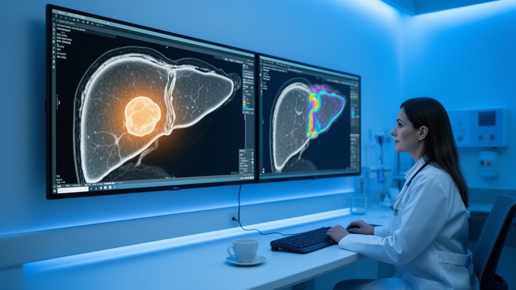
Liver cancer surgery is a high-stakes gamble. When surgeons cut out a hepatocellular carcinoma, the most common form of primary liver cancer, their success depends heavily on whether the tumor carries hidden features that mark it as aggressive. Now, a multicenter study from China suggests that artificial intelligence can read those features directly from MRI scans taken before the operation, potentially transforming how doctors plan treatment for one of the world’s deadliest cancers.
The research, published in BMC Medical Imaging by a team led by Wanjing Zheng and Dairong Cao of the First Affiliated Hospital of Fujian Medical University, focused on a clinical concept known as the MSE score. This score defines a hepatocellular carcinoma as high risk when it shows three ominous characteristics under the microscope or on imaging: microvascular or macrovascular invasion, meaning tumor cells have crept into blood vessels; satellite nodules, small daughter tumors lurking near the main mass; and a high Edmondson-Steiner grade, which reflects how wildly abnormal the cancer cells look. Any of these features portends a poor prognosis, because tumors that have already begun to invade vessels or seed new colonies tend to return after surgery, often with fatal consequences.
The problem is that none of these features can be reliably assessed before the tumor is removed. Pathologists need the resected specimen, and by then the surgical decisions have already been made. Surgeons operating blind on a high-risk tumor may underestimate how much liver to remove or how intensively to monitor the patient afterward. Preoperative liver biopsies can help, but they carry risks of bleeding and, in a twist that makes oncologists uneasy, may themselves seed tumor cells along the needle track. A noninvasive method to identify high-risk tumors before the first incision has therefore been a long-sought goal.
The Chinese team’s answer combines a specialized MRI contrast agent with a machine-learning approach called habitat imaging. The contrast agent, gadobenate dimeglumine, marketed as Gd-BOPTA and commonly known by the brand name MultiHance, has a peculiar property that makes it valuable for liver imaging. Unlike most gadolinium contrast agents, which are washed out of tissue quickly, a fraction of Gd-BOPTA is taken up by functioning hepatocytes and excreted into bile. This means that hours after injection, healthy liver tissue glows brightly on the scan while most tumors stand out as dark patches. The hepatobiliary phase images it produces carry rich information about how well tumor-adjacent liver cells are functioning and how the tumor interacts with its surroundings.
Habitat imaging, the study’s technical core, treats a tumor not as a single lump but as a patchwork of distinct biological microenvironments. The researchers divided every tumor voxel, the three-dimensional pixels of the MRI volume, into subregions using an algorithm called k-means clustering. Applied to ratio maps derived from the arterial phase, when blood flows into the tumor, and the hepatobiliary phase, when contrast clears through healthy liver cells, the clustering automatically partitions the tumor into habitats that may correspond to viable cancer, necrotic core, densely cellular zones, and invasive margins. Quantitative features extracted from each habitat describe texture, intensity distribution, and spatial patterns that the human eye cannot systematically evaluate. The idea is that aggressive tumors organize their internal architecture differently from indolent ones, and that this organization leaves fingerprints on dual-phase contrast-enhanced MRI.
To test the concept, the team assembled 583 patients from four hospitals. The largest center contributed 527 patients, split roughly seven to three into a training set used to build the model and an internal validation set used to tune expectations. Fifty-six patients from three other hospitals formed an external validation cohort, the crucial test of whether the model generalizes beyond the institution where it was born. From clinical records and scans, the researchers identified five independent predictors of high-risk disease using multivariable logistic regression. Elevated alpha-fetoprotein, a blood marker shed by many liver cancers, raised the odds of high-risk status more than fourfold. Two classic MRI signs also mattered: corona enhancement, a ring of contrast around the tumor suggesting microscopic invasion at its perimeter, raised odds more than fourfold, and peritumoral hypointensity, a dark halo around the tumor on hepatobiliary phase images, raised odds nearly elevenfold. Interestingly, non-rim arterial phase hyperenhancement, a pattern typically associated with typical, well-characterized tumors, was protective, reducing odds by about two-thirds.
But the strongest predictor by far was the habitat score derived from the machine-learned tumor subregions, which carried an odds ratio of 44.46, meaning patients with unfavorable habitat features faced dramatically higher odds of harboring an aggressive tumor. The researchers folded all five predictors into a nomogram, a graphical scoring tool that clinicians can use to combine multiple risk factors into a single probability estimate. The resulting Gd-BOPTA habitat fusion model achieved an area under the receiver operating characteristic curve, or AUC, of 0.844 in the training cohort, 0.797 in internal validation, and 0.820 in external validation. An AUC of 0.8 or above is generally considered acceptable-to-good discrimination in clinical prediction, and the fact that the external validation figure held up against the training numbers is the study’s most encouraging technical result, since many radiomics models collapse when moved to new hospitals with different scanners and protocols.
The model also passed a survival test. When the researchers grouped patients by the nomogram’s prediction and tracked recurrence-free survival after surgery, patients flagged as high risk relapsed far sooner than those flagged as low risk. Median recurrence-free survival was 18 months versus 56 months in the training cohort and 19 months versus 69 months in internal validation, differences that were statistically significant. In other words, the nomogram did more than mirror pathology; it anticipated the clinical course, suggesting that preoperative MRI habitat analysis captures genuine biological aggressiveness rather than merely correlating with it. Decision curve analysis, a method for weighing the benefits of acting on a prediction against the costs of being wrong, supported the model’s clinical utility across a plausible range of risk thresholds.
The implications, if the findings hold up, could be substantial. A surgeon armed with a preoperative risk score could widen resection margins around a predicted high-risk tumor, consider neoadjuvant or adjuvant therapy, prioritize the patient for liver transplantation where appropriate, or schedule more intensive surveillance for early recurrence. Because the tool builds on Gd-BOPTA MRI, which is already part of standard liver cancer workups in many centers, and packages its output into a simple nomogram, the barrier to clinical adoption is lower than for exotic imaging techniques requiring new hardware. The study’s authors are careful to note the caveats: the work is retrospective, meaning patients were identified from archives rather than followed prospectively, and the external validation cohort, though multi-institutional, was small at 56 patients. Large-scale prospective validation will be required before the nomogram earns a place in treatment guidelines. Still, the study adds to a growing body of evidence that the internal geography of a tumor, mapped computationally from routine scans, can reveal what a pathologist would otherwise need a scalpel to see. For patients with liver cancer, whose window for curative treatment is often heartbreakingly narrow, seeing the tumor’s true nature before the operation could mean the difference between a cure and a recurrence.
Subject of Research: Preoperative prediction of high-risk hepatocellular carcinoma using Gd-BOPTA MRI habitat imaging and a clinical nomogram
Article Title: Gd-BOPTA MRI-based nomogram incorporating habitat features for preoperative prediction of high-risk hepatocellular carcinoma: a multicenter retrospective study
Article References: Gd-BOPTA MRI-based nomogram incorporating habitat features for preoperative prediction of high-risk hepatocellular carcinoma: a multicenter retrospective study. (n.d.). https://doi.org/10.1186/s12880-026-02881-4
Image Credits: AI Generated
DOI: 10.1186/s12880-026-02881-4
Keywords: hepatocellular carcinoma, Gd-BOPTA MRI, habitat imaging, nomogram, microvascular invasion, radiomics, machine learning, recurrence-free survival, gadobenate dimeglumine, cancer imaging, prognosis, multicenter study
Cite Scienmag News
Ophelia Keating. (October 15, 2026). AI Reads Liver Scans to Spot Dangerous Tumors Before Surgery. Scienmag. https://scienmag.com/ai-reads-liver-scans-to-spot-dangerous-tumors-before-surgery/
Ophelia Keating. "AI Reads Liver Scans to Spot Dangerous Tumors Before Surgery." Scienmag, 15 October 2026, https://scienmag.com/ai-reads-liver-scans-to-spot-dangerous-tumors-before-surgery/. Accessed 15 October 2026.
Ophelia Keating. "AI Reads Liver Scans to Spot Dangerous Tumors Before Surgery." Scienmag. October 15, 2026. https://scienmag.com/ai-reads-liver-scans-to-spot-dangerous-tumors-before-surgery/


