


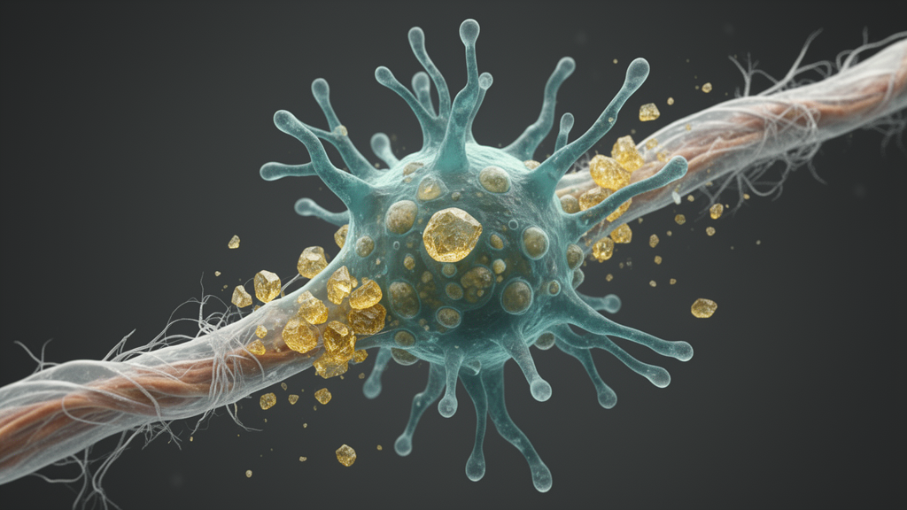
A distinct, pro-inflammatory microglial state marked by failing lysosomes and jammed cholesterol processing develops in the brains of mice with chronic progressive demyelination, and a drug that mimics the sterol desmosterol can partially reverse it, according to a study published in Nature Neuroscience. The work, led by Stefan Berghoff, Lena Spieth and Mikael Simons, compared genetic, toxic and autoimmune models of myelin loss and found that sustained myelin damage pushes microglia — the brain’s resident immune cells — into a state fundamentally different from the response seen after acute injury. In this chronic state, microglia sequester myelin-derived cholesterol inside their lysosomes instead of converting it into storage droplets, sustain production of the inflammatory chemokine CCL4, and lose the sterol-efflux program that normally allows them to hand off lipids for recycling. Restoring that program with a desmosterol mimetic improved motor performance and reduced myelin damage in a leukodystrophy model and eased disease in a mouse model of multiple sclerosis pathology.
The team’s central model was the Plp1Tg mouse, an animal carrying extra copies of the wild-type Plp1 gene and used to study Pelizaeus–Merzbacher disease, a human leukodystrophy caused by proteolipid protein overexpression. These mice develop initially normal myelin that then deteriorates progressively. By 12 weeks of age, labeling for myelin basic protein in the corpus callosum had dropped by roughly 30 percent, and by 24 weeks it had fallen by about 70 percent, a decline that tracked with worsening performance on the rotarod coordination test. Single-cell RNA sequencing of oligodendrocytes revealed a disease-associated cluster marked by the genes Serpina3n and C4b, alongside structural myelin abnormalities such as outfoldings and periaxonal vacuoles. The pathology was accompanied by reactive astrocytes, an influx of myeloid cells and CD3-positive T cells, particularly within white matter tracts, with flow cytometry detecting both CD4-positive and CD8-positive effector T cells as well as a substantial population of FoxP3-positive regulatory T cells.
A first question was whether this immune response was harmful or protective. The answer, from a series of depletion and drug experiments, was clearly protective. Depleting CD8-positive T cells with antibodies significantly impaired rotarod performance, and both CD4 and CD8 depletion increased abnormal myelin basic protein-positive swellings lacking nuclear labeling. Depleting microglia themselves with the CSF1R antagonist PLX5622 reduced IBA1-positive cell area by about 40 percent but made myelin pathology worse, most probably because the cells were no longer available to remove aberrant myelin. Anti-inflammatory and immune-modulatory treatments told a similar story: the Bruton’s tyrosine kinase inhibitor tolebrutinib and the JAK–STAT inhibitor baricitinib each significantly worsened rotarod performance, while interferon-1β and baricitinib treatment produced significantly increased myelin basic protein labeling and abnormal myelin swellings, and tolebrutinib caused only mild histological changes. The authors conclude that suppressing inflammation in this model impairs the immune-mediated clearance of damaged myelin rather than helping it.
To find out whether maladaptive microglial states coexisted with this necessary response, the researchers sequenced 57,141 single cells from Plp1Tg mice, mice fed the toxin cuprizone for six or twelve weeks, and controls. Microglia separated into six clusters: a homeostatic population, an interferon-responsive subset, and four disease-associated microglia subsets, DAM1 through DAM4. DAM1 was enriched for inflammatory and phagosome–lysosomal genes, including Ccl3, Ccl4, Ch25h and Lpl, while DAM4 carried antigen-presentation and lipid-trafficking programs. In Plp1Tg mice, the DAM1 population expanded to roughly 23 percent of all microglia, and its abundance rose with disease stage in pseudotime trajectory analysis. Immunohistochemistry showed that about 30 percent of microglia were CCL4-positive and largely distinct from MHCII-positive cells. Critically, co-labeling revealed myelin basic protein accumulating inside the CD68-positive lysosomes of CCL4-positive microglia, and electron microscopy showed electron-dense lysosomal material, intralysosomal myelin stacks and cholesterol crystals within these cells. Blocking CCL4 signaling with the CCR5 antagonist maraviroc, however, did not significantly alter rotarod performance or histological measures, suggesting CCL4 marks the state rather than simply driving it.
The most striking metabolic contrast emerged when the team compared how microglia handle myelin lipids in acute versus chronic injury. After six weeks of cuprizone, which induces oligodendrocyte death and demyelination, more than half of IBA1-positive microglia contained PLIN2-positive lipid droplets — the hallmark of so-called foamy phagocytes that buffer excess cholesterol by esterifying it. In 24-week-old Plp1Tg mice, such droplets were almost entirely absent from microglia. Instead, PLIN2-positive lipid droplets accumulated in GFAP-positive astrocytes, a shift also seen after twelve weeks of cuprizone. Untargeted lipidomics of isolated microglia confirmed the divergence: both models showed elevated myelin-associated hexosylceramides, but Plp1Tg microglia additionally accumulated very long-chain ceramides and bis(monoacylglycerol)phosphate, a lysosomal lipid linked to impaired lipid handling, while showing reduced cholesterol esters. The authors interpret this as impaired cholesterol esterification and lysosomal lipid handling during chronic progressive myelin pathology.
Tracing experiments pinned the defect to intracellular cholesterol trafficking. When a fluorescent cholesterol derivative was injected into the corpus callosum, 78 percent of the label localized to PLIN2-positive lipid droplets within microglia of wild-type mice recovering from a lysolecithin lesion, whereas in Plp1Tg mice 98 percent of the tracer accumulated in LAMP1-positive endosomes and lysosomes. Genetic tests supported the same mechanism. Microglia-specific deletion of NPC1, the transporter that exports free cholesterol from late endosomes and lysosomes, reproduced the entire phenotype after focal demyelination: increased myelin basic protein inside microglia, a complete absence of PLIN2-positive droplets, elevated CCL4-positive cells and crystalline lysosomal material on electron microscopy. Deleting SOAT1, the enzyme that esterifies cholesterol for droplet storage, produced the same triad. In primary microglia cultures, myelin debris upregulated lysosomal and sterol genes including Ccl4, and pharmacological inhibition of SOAT1 or NPC1 increased the number of CCL4-positive cells while suppressing lipid droplet formation.
Transcriptional profiling explained why the cells cannot escape. Compared with microglia from six-week cuprizone-treated mice, Plp1Tg microglia — and the CCL4-high DAM1 subset in particular — showed reduced expression of the lipid-efflux genes Apoe, Abca1 and Abcg1, together with increased expression of cholesterol-synthesis genes, including Dhcr24, which encodes 24-dehydrocholesterol reductase, the terminal enzyme that converts desmosterol into cholesterol. Gas chromatography–tandem mass spectrometry confirmed that desmosterol rose in the corpus callosum after six weeks of cuprizone but not in Plp1Tg tissue or after twelve weeks of cuprizone. Because desmosterol is an endogenous ligand for the liver X receptors, which promote lipid efflux and limit inflammatory responses, the authors conclude that chronic myelin pathology uncouples sterol synthesis from sterol efflux, denying microglia the signal they need to switch into a regenerative, lipid-exporting mode.
The therapeutic test followed directly from this mechanism. The researchers treated Plp1Tg mice from two to five months of age with DMHCA, a compound that acts both as a liver X receptor agonist and as a DHCR24 inhibitor, allowing desmosterol to accumulate. DMHCA improved rotarod performance, reduced myelin loss without producing myelin swellings, increased CAII-positive oligodendrocyte numbers and reduced IBA1-positive cell counts. It also lowered the number of CCL4-positive microglia and shrank the CD68-positive lysosomal area. SH42, a selective DHCR24 inhibitor lacking liver X receptor agonist activity, produced similar but weaker effects, while GW3965, a direct nonsteroidal liver X receptor agonist that does not inhibit DHCR24, had no detectable effect — indicating that the DHCR24–desmosterol pathway, not generic receptor activation, is what matters. In ex vivo slice cultures, Plp1Tg microglia took up more myelin than wild-type cells, and DMHCA reduced microglial numbers, lysosomal area and intracellular myelin content. Notably, supplementing the diet with the sterol squalene supported developmental myelination but did not ameliorate pathology once demyelination had begun.
The findings extended to inflammatory demyelination more broadly. Using RNAscope on human autopsy tissue, the team found that roughly 75 percent of CD68-positive phagocytes in chronic active multiple sclerosis lesions expressed CCL4, compared with about 13 percent in non-MS control white matter, and around 35 percent of these double-positive cells showed strong autofluorescence suggestive of lysosomal storage material. In the experimental autoimmune encephalomyelitis model, DMHCA treatment started at symptomatic onset improved disease severity, reduced lesion size and number, increased myelin basic protein area, lowered CD4-positive T cell infiltration and reduced CCL4-positive microglia and Ccl4 transcripts in isolated CD11b-positive cells. These human and EAE data are correlative and preclinical respectively, and the maraviroc result shows that targeting a single CCL4 downstream receptor is insufficient, but they support the idea that the CCL4-positive, lysosome-clogged phagocyte state is relevant beyond the Plp1Tg model.
The study proposes a two-mode model of cholesterol clearance: after acute injury, microglia move lysosomal cholesterol to the endoplasmic reticulum and store it transiently as esterified lipid droplets, whereas in chronic pathology that route fails, astrocytes take over droplet storage, and microglia remain locked in a pro-inflammatory, myelin-reactive state that resembles the phagocyte phenotypes of lysosomal storage disorders. Because myelin is a dense, cholesterol-rich membrane that is difficult to degrade, and because lingering myelin debris blocks oligodendrocyte precursor cell recruitment and differentiation, efficient clearance is essential for repair. The connection between lysosomal function, sterol metabolism and demyelination identifies the DHCR24–desmosterol axis as a potential therapeutic target for chronic progressive demyelinating disorders, although the work so far rests on mouse models, small cohorts and one preclinical drug paradigm, and human validation remains to be established.
Subject of Research: Microglial lysosomal dysfunction and sterol metabolism in chronic progressive demyelination
Article Title: Chronic progressive demyelination induces a microglial state with lysosomal dysfunction
Article References: Berghoff, S. A., Spieth, L., Vitale, S., Schlaphoff, L., Zirngibl, M., Junker, N., Dean, A.-C., Liu, L., Damkou, A., Roth, S., Hurler, C.-A., Hosang, L., Perez Anderson, K., Besson-Girard, S., Reich, M., Feiten, A., Huber, L., Müller, C., Kislinger, G., … Simons, M. (2026). Chronic progressive demyelination induces a microglial state with lysosomal dysfunction. Nature Neuroscience. https://doi.org/10.1038/s41593-026-02489-5
Image Credits: AI Generated
DOI: 10.1038/s41593-026-02489-5
Keywords: microglia, demyelination, lysosomes, cholesterol metabolism, desmosterol, DHCR24, CCL4, multiple sclerosis, leukodystrophy, Plp1Tg mouse model, lipid droplets, myelin clearance
Cite Scienmag News
Ophelia Keating. (October 26, 2026). Chronic Demyelination Traps Microglia in a Lysosomal Cholesterol State. Scienmag. https://scienmag.com/chronic-demyelination-traps-microglia-in-a-lysosomal-cholesterol-state/
Ophelia Keating. "Chronic Demyelination Traps Microglia in a Lysosomal Cholesterol State." Scienmag, 26 October 2026, https://scienmag.com/chronic-demyelination-traps-microglia-in-a-lysosomal-cholesterol-state/. Accessed 26 October 2026.
Ophelia Keating. "Chronic Demyelination Traps Microglia in a Lysosomal Cholesterol State." Scienmag. October 26, 2026. https://scienmag.com/chronic-demyelination-traps-microglia-in-a-lysosomal-cholesterol-state/


