


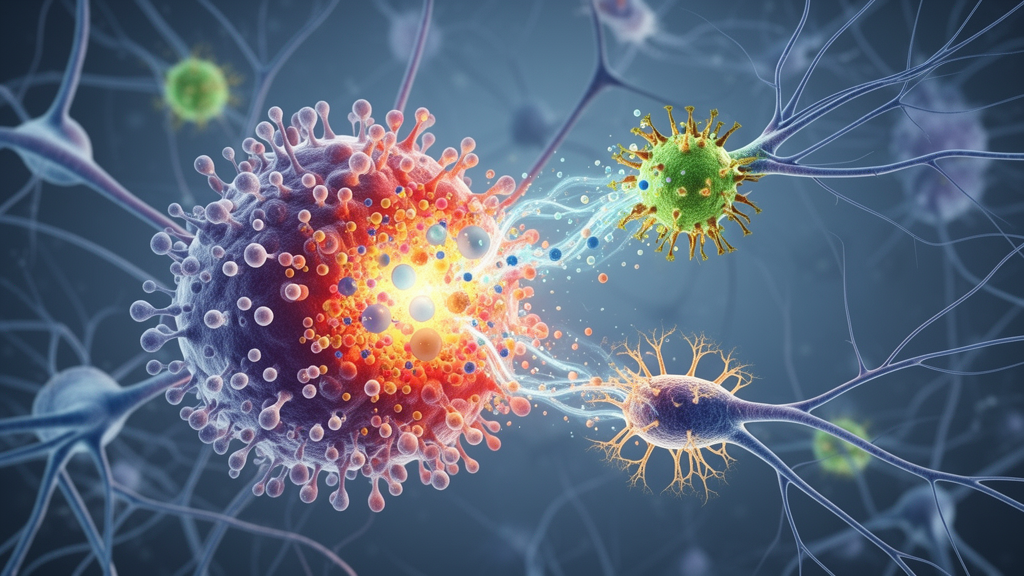
Vestibular migraine, a disorder marked by recurrent vertigo often accompanied by headache, nausea, and vomiting, affects roughly 2.7 percent of adults according to a nationwide study applying the International Classification of Headache Disorders, Third Edition criteria. Despite its prevalence, the underlying mechanisms remain incompletely understood, and no specific biomarkers exist for diagnosis, which currently relies on clinical history and symptom evaluation. A new review published in Frontiers in Neuroscience argues that a promising way forward lies in an underappreciated cellular conversation: the bidirectional signaling between microglia and astrocytes, the central nervous system’s principal immune-regulatory cells, which may help sustain the abnormal brain-state known as central sensitization that underlies many vestibular migraine symptoms.
Central sensitization describes structural, functional, and neurochemical changes in the central nervous system that heighten sensitivity to pain and other sensory inputs. It is considered a key mechanism in migraine chronification, and the review’s authors, based at Henan University of Traditional Chinese Medicine and affiliated clinical centers, contend that it may similarly drive both the headache and vertigo of vestibular migraine. Microglia serve as the resident immune cells of the brain, while astrocytes, the most abundant glial cells, regulate neurotransmission, inflammation, neurogenesis, and synaptic plasticity. When these two cell types exchange inflammatory signals, the review suggests, they can amplify neuroinflammation and neuronal excitability in ways that promote and maintain sensitization.
The authors are careful to distinguish between loose functional relationships and what they call true crosstalk. In their framework, crosstalk requires experimental evidence that a signal produced or regulated by one cell type influences the molecular or functional response of the other, with both cell types clearly identified. Such evidence may include a temporal or functional association, a ligand–receptor pairing, changes in downstream signaling, or support from pharmacological or genetic manipulation. Bidirectional communication is not required; where evidence supports signaling in only one direction, the direction is stated explicitly. They also caution that simple binary classifications of microglia into M1 and M2 states and astrocytes into A1 and A2 phenotypes are inadequate, since glial responses are shaped by stimulus type and disease stage into dynamic, heterogeneous states.
One of the better-supported signaling routes runs from microglia to astrocytes. In experimental studies, activated microglia release the cytokines IL-1α, TNF-α, and C1q, which induce reactive changes in astrocytes. Microglia-derived TNF-α promotes inflammatory signaling in astrocytes through nuclear translocation of the transcription factor NF-κB. Timing experiments with lipopolysaccharide stimulation show a sequential pattern: pro-inflammatory cytokine expression in microglia peaks two to four hours after stimulation, while cytokine and chemokine induction in astrocytes peaks later, at twelve hours, consistent with microglia detecting stimuli early and shaping the subsequent astrocyte response. In a migraine model induced by dural infusion of inflammatory soup, interleukin-18 was localized mainly in microglia while its receptor was found mainly on astrocytes, and blocking this IL-18 signaling reduced astrocyte reactivity and migraine-like pain hypersensitivity, providing relatively direct cell-specific evidence of microglia-to-astrocyte transmission.
Signaling in the reverse direction is also documented. TNF-α-stimulated astrocytes can express and release the chemokine CCL2, which modulates microglial inflammatory responses through the CCR2 receptor; inhibiting CCR2 attenuates the effect. In a rat model of chronic migraine, increased CCL2 in the trigeminal nucleus caudalis was mainly colocalized with astrocytes, while CCR2 was associated with microglia, and recombinant CCL2 enhanced NLRP3-related inflammatory responses in microglia alongside elevations of the sensitization markers c-Fos and phosphorylated ERK. In a neuropathic pain study, astrocyte-derived CSF1 acted on CSF1R receptors on microglia, promoting microglial responses and brain-derived neurotrophic factor production that increased spinal neuron excitability; blocking this pathway attenuated central sensitization and allodynia. The CXCL12–CXCR4 axis offers a further candidate route from astrocytes to microglia.
Within vestibular migraine models specifically, evidence is emerging but still limited. Animal studies have found significantly increased IL-1β levels in both the vestibular nucleus and the trigeminal nucleus caudalis, along with reactive glial changes in the vestibular nucleus–trigeminal nucleus region. One recent study reported increased expression of the ion channel TRPV2 in microglia of the trigeminal nucleus caudalis in a vestibular migraine mouse model, accompanied by enhanced NLRP3 inflammasome activity and elevated pro-inflammatory mediators; in vitro experiments confirmed functional TRPV2 activity, and its inhibition reduced inflammasome activation, implicating a TRPV2–calcium–NLRP3 signaling axis. Another study in vestibular migraine models found that the CXCL10/CXCR3 pathway, acting through PI3K/AKT signaling, was associated with increased IL-1β, IL-6, and TNF-α in trigeminal-related regions.
The downstream consequences of these glial signals converge on the trigeminovascular system, the network whose activation releases vasoactive neuropeptides such as calcitonin gene-related peptide, promoting vasodilation, sterile inflammation, and pain signal transmission into the central nervous system. In migraine models, reactive microglia and astrocytes appear within the trigeminal nucleus caudalis, and the duration of their reactivity correlates with trigeminovascular sensitization. Microglial P2X4 receptor signaling promotes BDNF release, raising neuronal excitability in the trigeminal nucleus, while inhibition of the microglial NLRP3–IL-1β pathway reduces markers of central sensitization including p-ERK, c-Fos, and CGRP and alleviates hyperalgesia. On the astrocyte side, reduced expression of the glutamate transporter EAAT2 in chronic migraine rats impaired glutamate homeostasis, and upregulating EAAT2 reduced NMDA receptor-related signaling and abnormal synaptic plasticity, suggesting astrocytes tune trigeminal neuron excitability through glutamate clearance.
Glutamate dysregulation emerges as a central theme linking glial crosstalk to symptoms. Reactive microglia can release glutamate directly and can stimulate astrocyte glutamate release via ATP-driven P2Y1 receptor signaling, while microglial reactivity disrupts the astrocyte-dependent glutamate–glutamine cycle that normally clears synaptic glutamate and prevents excitotoxic damage. Microglia can even prune astrocytic processes, reducing their coverage of neurons and promoting activation of extrasynaptic NMDA receptors. Excess glutamate relieves the magnesium block of NMDA receptor channels, allowing calcium influx and intracellular cascades that sustain sensitization. Notably, magnetic resonance measurements in patients during vestibular migraine attacks have found elevated levels of the glutamate–glutamine complex in the bilateral thalamus, hinting that this cycle is disturbed in the human condition, although the review stresses that most mechanistic detail comes from animal work.
The review also sketches an upstream pathway from the body’s periphery. Peripheral inflammation can induce widespread glial and cytokine responses in the central nervous system, with the microglial response preceding the astrocytic one, and inflammatory factors such as TNF-α and IL-1β can disrupt blood–brain barrier tight junctions. In one chronic migraine model, the cytokine IL-17A penetrated the blood–brain barrier into the medulla and activated the trigeminal nucleus caudalis through microglia-mediated neuroinflammation. Separately, migraine-related neuropeptides including CGRP may enter the inner ear through a disrupted blood–labyrinth barrier; animal studies show CGRP injection alters auditory and vestibular evoked potentials and induces behaviors resembling the phonophobia and postural instability of patients, and repeated nitroglycerin treatment raised CGRP and its receptors in cochlear outer hair cells, causing mitochondrial stress, DNA leakage, and cell death that CGRP receptor antagonists reduced. Trigeminal fibers innervate the labyrinthine vasculature, and glutamatergic projections run from the trigeminal nucleus caudalis to the vestibular nucleus, silencing which markedly improves nitroglycerin-induced vestibular dysfunction in rats.
Synthesizing this evidence, the authors propose a pathological sequence for vestibular migraine running from peripheral trigger, through abnormal sensory input or neuroimmune transmission, to glial response, to central sensitization, with glial crosstalk then amplifying neuroinflammation in a bidirectional feedback loop. They acknowledge substantial limitations: most mechanistic insights derive from animal models of migraine, chronic migraine, or pain rather than vestibular migraine itself, direct evidence for microglia–astrocyte crosstalk within the trigeminovascular system remains sparse, and no therapies that explicitly target glial crosstalk have yet been validated. The authors call for human-centered approaches, including neuroimaging, cerebrospinal fluid analysis, single-cell sequencing, and spatial transcriptomics, along with better animal models that reproduce both headache-like behaviors and vestibular dysfunction, before these mechanisms can be translated into biomarkers or treatments for patients.
Subject of Research: Microglia–astrocyte signaling crosstalk in central sensitization of vestibular migraine
Article Title: Research progress on the mechanism of microglia–astrocyte signal crosstalk in central sensitization of vestibular migraine
Article References: Jin, T., Wang, W., Qin, J., Liu, Y., Fan, X., Zhang, D., & Zhang, H. (2026). Research progress on the mechanism of microglia–astrocyte signal crosstalk in central sensitization of vestibular migraine. Frontiers in Neuroscience, 20, Article 1844932. https://doi.org/10.3389/fnins.2026.1844932
Image Credits: AI Generated
DOI: 10.3389/fnins.2026.1844932
Keywords: vestibular migraine, microglia, astrocytes, central sensitization, neuroinflammation, trigeminovascular system, glutamate, CGRP, NLRP3 inflammasome, blood-brain barrier, glial crosstalk, trigeminal nucleus caudalis
Cite Scienmag News
Cassandra Pierce. (October 23, 2026). Glial Cell Crosstalk May Drive Central Sensitization in Vestibular Migraine, Review Finds. Scienmag. https://scienmag.com/glial-cell-crosstalk-may-drive-central-sensitization-in-vestibular-migraine-review-finds/
Cassandra Pierce. "Glial Cell Crosstalk May Drive Central Sensitization in Vestibular Migraine, Review Finds." Scienmag, 23 October 2026, https://scienmag.com/glial-cell-crosstalk-may-drive-central-sensitization-in-vestibular-migraine-review-finds/. Accessed 23 October 2026.
Cassandra Pierce. "Glial Cell Crosstalk May Drive Central Sensitization in Vestibular Migraine, Review Finds." Scienmag. October 23, 2026. https://scienmag.com/glial-cell-crosstalk-may-drive-central-sensitization-in-vestibular-migraine-review-finds/


