


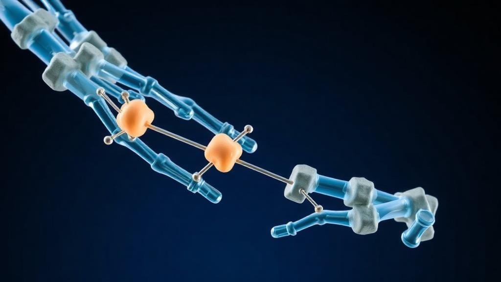
Bone is not the inert scaffold it appears to be. It is a living tissue locked in a continuous tug-of-war between cells that build new bone and cells that dismantle old bone, and when that balance slips, the result is osteoporosis, osteoarthritis, or failed fracture healing. A new review published in BMC Medicine argues that scientists have been watching only half of this regulatory drama. The authors, led by Yunpeng Zou and Ronghan Liu of Shandong Second Medical University and collaborating institutions, contend that post-translational modifications, the chemical tags attached to proteins after they are made, form a hidden control layer that shapes how bone cells respond to the classical signaling pathways long considered the master regulators of skeletal biology.
Post-translational modifications, or PTMs, are biochemical edits applied to proteins after translation. Phosphorylation, ubiquitination, acetylation, methylation, SUMOylation, O-GlcNAcylation, and poly-ADP-ribosylation each alter a protein’s behavior in distinct ways. A phosphate group can switch an enzyme on or off. A ubiquitin chain can mark a transcription factor for destruction. An acetyl group can change where a protein sits inside the cell or whom it binds to. The review’s central claim is that these modifications do not merely fine-tune bone biology; they act as a networked control system that gates, amplifies, and dampens the signals flowing through pathways such as Wnt, BMP, RANKL, Notch, and Hedgehog, which orchestrate the activity of osteoblasts, osteoclasts, and osteocytes.
The authors organize the evidence around three conceptual pillars. The first is bidirectionality: PTMs and classical signaling pathways do not operate in a one-way hierarchy but feed back into each other. Signaling cascades recruit enzymes that modify target proteins, while those modified proteins in turn alter the intensity, duration, and spatial distribution of the signals themselves. The second pillar concerns functional modes. Some PTM events act as gates, holding a pathway silent until a specific cue arrives. Others amplify a weak signal into a robust cellular response, or dampen an overactive pathway to prevent runaway bone resorption. The third pillar is spatiotemporal regulation: the same modification can have different consequences depending on which cell type carries it, in which subcellular compartment it occurs, and at which stage of development, aging, or disease it appears.
Osteoblasts, the bone-forming cells derived from mesenchymal stem cells, illustrate the stakes. Their differentiation depends on transcription factors such as RUNX2 and Osterix, whose activity and stability are controlled by phosphorylation and ubiquitination. Glycogen synthase kinase 3 beta, a well-known kinase, phosphorylates beta-catenin in the Wnt pathway and tags it for degradation, thereby suppressing osteoblast differentiation. When GSK3β itself is phosphorylated at an inhibitory site, beta-catenin accumulates and drives bone formation. The review emphasizes that such regulatory loops are not isolated curiosities but recurring motifs: the same kinase or ligase can be repurposed by different PTM events to produce opposite outcomes depending on context.
Osteoclasts, the bone-resorbing cells, are governed by an equally dense PTM web. The RANKL signaling cascade, which drives osteoclast differentiation and activation, depends on phosphorylation events that propagate signals from the cell surface to the transcription factor NFATc1, the master regulator of osteoclast identity. Ubiquitination and deubiquitination of signaling intermediates determine whether the response is sustained or extinguished. Cathepsin K, the protease that dissolves bone matrix, is itself subject to regulatory processing. The review highlights that dysregulated PTM events in osteoclasts contribute directly to pathological bone loss in osteoporosis and inflammatory conditions such as rheumatoid arthritis, where tumor necrosis factor alpha and interleukin-6 reshape the modification landscape of signaling proteins.
Osteocytes, the embedded mechanosensory cells that make up the vast majority of bone cells, add another layer of complexity. These cells sense mechanical loading and orchestrate remodeling by secreting molecules such as sclerostin and RANKL. The review notes that PTMs regulate osteocyte function through modifications of proteins involved in mechanotransduction, the process by which physical force is converted into biochemical signals. O-GlcNAcylation, the attachment of N-acetylglucosamine sugars to proteins by the enzyme OGT and their removal by OGA, emerges as a nutrient-sensitive modification that links cellular metabolism to bone cell behavior. Histone acetylation and methylation, mediated by histone acetyltransferases and deacetylases, control the accessibility of genes required for osteocyte survival and communication within the bone matrix.
The pathological implications are substantial. The review connects dysregulated PTMs to osteoporosis, osteoarthritis, metabolic bone diseases, inflammatory bone loss, and impaired fracture repair. In osteoarthritis, modifications of cartilage and subchondral bone proteins alter the balance between matrix degradation and synthesis. In senescence-associated conditions, PTMs influence the secretory phenotype of aging skeletal cells, which promotes local inflammation and tissue degeneration. Poly-ADP-ribosylation, a modification involved in DNA damage responses, appears in the context of oxidative stress and cell death in bone cells. Peptidylarginine deiminase enzymes, which convert arginine residues to citrulline, are implicated in inflammatory joint disease and may influence bone erosion. Each of these connections points to PTM enzymes as potential therapeutic targets, though the review is careful to note that causality remains unproven for many site-specific events.
That caution is one of the review’s most valuable contributions. The authors acknowledge that evidence is uneven across PTM types and skeletal cell populations. Phosphorylation is well characterized in osteoblast and osteoclast signaling, but less-studied modifications such as SUMOylation, neddylation, and lactylation remain poorly mapped in bone. Many reported PTM events are correlational rather than causal; showing that a modification is present in a disease state does not prove it drives the pathology. Distinguishing which of the thousands of possible modification events actually control bone homeostasis requires targeted mutagenesis of specific modification sites, cell-type-specific deletion of modifying enzymes, and quantitative proteomics that can track modification dynamics over time and across compartments.
Emerging technologies are beginning to close these gaps. Quantitative PTM proteomics now allows researchers to measure thousands of modification events simultaneously and compare their abundance between healthy and diseased bone. Single-cell RNA sequencing and spatial transcriptomics reveal which cell populations carry which modification machinery, resolving the heterogeneity that bulk assays obscure. Multi-omics integration, combining proteomics, transcriptomics, epigenomics, and metabolomics, offers a systems-level view of how PTM networks interact with metabolic state and gene expression. The review argues that these tools, combined with classical genetics and pharmacology, will be essential for identifying which PTM events are true regulatory nodes rather than passive consequences of cellular state.
The broader significance of the review lies in its reframing of bone biology as a networked control problem rather than a linear cascade of pathways. Just as engineers design control systems with sensors, actuators, and feedback loops, skeletal cells deploy PTMs as molecular switches that sense context and adjust signal flow accordingly. Understanding this architecture could transform how researchers approach bone diseases, shifting attention from individual pathways to the modification events that tune them. For patients with osteoporosis or non-healing fractures, the promise is that targeting the right PTM node could restore balance to the remodeling cycle with greater precision than current therapies, which broadly suppress bone resorption or stimulate formation without addressing the underlying regulatory logic. The review does not claim to have found those targets yet, but it provides a roadmap for where to look and a framework for evaluating what is found.
Subject of Research: Post-translational modifications regulating osteoblast, osteoclast, and osteocyte signaling in bone homeostasis and disease
Article Title: Networked control of bone homeostasis: interfacing post-translational modifications with cellular signaling
Article References: Zou, Y., Kang, J., Li, Z., Niu, J., Wang, Z., Wang, S., Han, X., Ning, B., & Liu, R. (2026). Networked control of bone homeostasis: interfacing post-translational modifications with cellular signaling. BMC Medicine. https://doi.org/10.1186/s12916-026-05308-7
Image Credits: AI Generated
DOI: 10.1186/s12916-026-05308-7
Keywords: bone homeostasis, post-translational modifications, osteoblasts, osteoclasts, osteocytes, bone remodeling, osteoporosis, osteoarthritis, phosphorylation, ubiquitination, O-GlcNAcylation, cellular signaling
Cite Scienmag News
Ophelia Keating. (October 16, 2026). Hidden Protein Switches That Keep Bones in Balance May Reshape Osteoporosis Research. Scienmag. https://scienmag.com/hidden-protein-switches-that-keep-bones-in-balance-may-reshape-osteoporosis-research/
Ophelia Keating. "Hidden Protein Switches That Keep Bones in Balance May Reshape Osteoporosis Research." Scienmag, 16 October 2026, https://scienmag.com/hidden-protein-switches-that-keep-bones-in-balance-may-reshape-osteoporosis-research/. Accessed 16 October 2026.
Ophelia Keating. "Hidden Protein Switches That Keep Bones in Balance May Reshape Osteoporosis Research." Scienmag. October 16, 2026. https://scienmag.com/hidden-protein-switches-that-keep-bones-in-balance-may-reshape-osteoporosis-research/


