


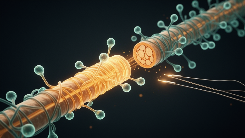
Multiple sclerosis has long been treated as a disease of immune attack, and the drugs that suppress that attack have transformed lives. Yet the myelin sheaths stripped from axons during each demyelinating episode rarely return to full integrity, and the neurological disability that accumulates over decades is the direct consequence of that failed repair. A new perspective article published in Frontiers in Molecular Neuroscience argues that the field has been asking too narrow a question. Rather than viewing remyelination failure solely as a problem of oligodendrocyte precursor cell differentiation, researchers Thomas Jonathan Scheinok, Dimitri De Bundel, Tim Vanmierlo and Jeroen Van Schependom propose that the hostile, inflammatory and age-related microenvironment surrounding MS lesions is equally responsible, and that only a combination strategy targeting both intrinsic and extrinsic mechanisms is likely to succeed where single-target drugs have repeatedly fallen short.
The biological case is compelling. In the early disease course, endogenous remyelination by oligodendrocyte precursor cells, and to a lesser extent by surviving oligodendrocytes, often restores the myelin sheath to near-complete integrity. But with ageing, this repair mechanism deteriorates. Microglia that accumulate in demyelinated lesions of aged animals adopt a senescence-associated secretory phenotype, chronically upregulating pro-inflammatory mediators while losing phagocytic efficiency. Myelin debris, which contains potent inhibitors of oligodendrocyte differentiation such as myelin-associated glycoprotein and Nogo-A, therefore lingers in the lesion. Simultaneously, pro-inflammatory cytokines including IL-1β and TNF-α, along with reactive oxygen species, create a hostile milieu for oligodendrocyte development. Astrocytes undergo a parallel decline: the transition from reactive to pro-regenerative phenotypes becomes impaired with age, growth factor production falls, and reactive astrocytes secrete extracellular matrix proteins that form glial scars and release neurotoxins.
Ageing of the axon-myelin unit itself compounds the problem. Myelin progressively degenerates in the ageing brain, and the resulting debris accumulates in microglial lysosomes, increasing the clearance burden on microglia that are already less efficient. Myelinated axons depend on oligodendrocytes for metabolic support, receiving glycolytic products such as glucose and lactate to maintain axonal integrity. This support is reduced in demyelinated or thinly remyelinated axons while the energy cost of conduction rises. Furthermore, ageing white matter is more vulnerable to metabolic and ischemic stress, as demonstrated by studies showing that optic nerves from older mice suffer more severe injury during oxygen-glucose deprivation than those of young mice, a finding particularly relevant to older people with MS in whom vascular comorbidities accumulate.
Against this backdrop, the clinical track record of pro-remyelinating pharmacotherapy is sobering. Clemastine, an antihistaminic drug with antimuscarinic properties, met its primary electrophysiological endpoint in the ReBUILD trial, shortening visual evoked potential P100 latency by 1.7 milliseconds per eye, and subsequent myelin water imaging provided convergent evidence of increased myelin content in the corpus callosum. Nevertheless, the magnitude and clinical relevance of that latency change remain uncertain, and a clemastine study in progressive MS was halted after an unexpected acceleration of disability. PIPE-307, a more selective M1 receptor antagonist designed to address the unfavourable side-effect profile, was well tolerated in the phase 2 VISTA trial but failed to meet its primary endpoint of improvement in low-contrast letter acuity. Opicinumab, an anti-LINGO-1 antibody, did not delay disease progression in people with MS despite being tested as add-on therapy to anti-inflammatory disease-modifying treatments. Bexarotene produced a negative primary MRI endpoint with poor tolerability in CCMR One.
The authors contend that these translational failures stem partly from a mechanistic blind spot: most investigated remyelinating pharmaceuticals act intrinsically on oligodendrocyte precursor cells, promoting lineage progression, while insufficiently addressing the non-permissive inflammatory, senescent and extracellular-matrix-rich microenvironment of MS lesions. Preclinical evidence supports this interpretation. Combining an anti-CD20 compound with clemastine fumarate significantly enhanced remyelination, reduced inflammatory activity and improved functional recovery compared to either treatment alone in an experimental autoimmune encephalomyelitis model. Similarly, tuftsin, an immunomodulatory tetrapeptide that induces an anti-inflammatory shift in microglia and macrophages, combined with benztropine, an anticholinergic drug with remyelinating capacity, produced comparable benefits. Molecules targeting glial inflammatory signalling, such as PPARγ agonists and IL-4, enhanced oligodendrocyte development and remyelination, while the Toll-like receptor 3 agonist Poly-IC increased interleukin-33 production in oligodendrocyte precursor cells, stimulating myelin gene transcription and cell maturation. Remodelling the extracellular matrix with inhibitors of chondroitin sulfate proteoglycan synthesis led to enhanced recruitment and differentiation of oligodendrocyte precursor cells, followed by remyelination.
Into this framework, the authors introduce non-invasive brain stimulation as a mechanistically distinct therapeutic strategy. Techniques such as repetitive transcranial magnetic stimulation, transcranial direct current stimulation and transcranial alternating current stimulation modulate neural network activity without surgery, and mounting evidence from healthy and demyelinating animal models suggests that modulating neuronal activity can positively affect oligodendrocyte proliferation, differentiation and maturation, as well as subsequent remyelination. Optogenetic stimulation of a subset of neurons demonstrates the neuronal activity-dependent maturation of oligodendrocytes, while blocking synaptic release impairs central nervous system myelination. In an ethidium bromide myelin lesion model, tetrodotoxin, an action potential-inhibiting toxin, impaired oligodendrocyte development and axon remyelination, revealing the importance of axonal signalling in repair. Oligodendrocyte precursor cells express various neurotransmitter receptors, and in vitro experiments have found that silencing those receptors disrupts cell development, whereas stimulating them can effectively boost oligodendrogenesis. Interestingly, the pattern of neuronal activity seems to influence precursor cell behaviour, with high-frequency stimulation boosting proliferation while low-frequency electrical activity promotes differentiation.
Crucially, activity-dependent effects on remyelination are unlikely to be mediated exclusively through direct neuron-oligodendrocyte precursor cell communication. Astrocytes and microglia also express receptors for neurotransmitters and neuromodulators, allowing them to respond directly to extracellular signalling changes induced by neuronal activity or stimulation. Neuronal depolarization triggers potassium release at nodes of Ranvier, which is sensed by microglia via the potassium channel THIK-1, inducing a shift towards pro-regenerative microglial transcriptional states necessary for myelin debris clearance. In astrocytes, neurotransmitter and neuromodulator signalling can induce intracellular calcium responses that influence astrocytic phenotype and function; transcranial direct current stimulation-induced astrocytic calcium signalling has been linked to noradrenergic alpha-1 receptor mechanisms in the healthy mouse cortex. Preclinical studies in demyelinating models strengthen the rationale: low-intensity repetitive transcranial magnetic stimulation delivered as intermittent theta burst stimulation enhanced remyelination by increasing the number of myelin internodes elaborated by new oligodendrocytes and by supporting the myelinating capacity of surviving oligodendrocytes, while 10 Hz transcranial alternating current stimulation enhanced oligodendrogenesis and myelin repair following cuprizone-induced demyelination.
Evidence for stimulation-induced glial modulation, in contrast, comes largely from models other than demyelination. Repetitive transcranial magnetic stimulation at 10 Hz shows a remarkable capacity to inhibit astrocyte polarization towards a pro-inflammatory phenotype while promoting a shift towards pro-regenerative states in an ischemic rat model. High-frequency stimulation can shift the microglial phenotype towards regeneration via the JAK2/STAT3 pathway, attenuating white matter lesions in a post-stroke rat model. A systematic review confirms that, across multiple disease models, high-frequency stimulation consistently decreases markers of astrocyte reactivity and attenuates microglial pro-inflammatory activation. Importantly, these glial modulatory effects appear to be context-dependent and more pronounced in injured than in healthy brain tissue, suggesting that the therapeutic window may be particularly relevant in the pathological context of MS. However, the authors caution that the injury in these models is ischemic rather than immune-mediated, and whether the glial mechanisms described in ischemic and post-stroke models also apply to MS lesions remains to be demonstrated.
The authors argue that pharmacological agents and stimulation may influence different processes of the remyelination cascade, making them genuinely complementary rather than redundant. Clemastine and PIPE-307 target the muscarinic M1 receptor-mediated inhibition of oligodendrocyte precursor cell differentiation, whereas thyromimetics and bexarotene activate myelin gene expression. Metformin, on the other hand, seems to restore the responsiveness of aged precursor cells to differentiation cues. Stimulation, by contrast, mainly influences the environment in which precursor cells must differentiate as well as the axonal signals that guide myelination. First, stimulation may shift the intrinsic states of microglia and astrocytes towards regeneration and support myelin debris clearance. Second, neuronal activity influences which axons get myelinated and how many internodes are formed by new oligodendrocytes, so a drug that increases differentiation may benefit from a supplementary activity-dependent signal that directs these cells towards myelin sheath generation. Third, metformin may intrinsically act on the metabolism of aged precursor cells, whereas stimulation could improve the toxic extrinsic glial microenvironment.
Yet the authors also warn that some mechanisms of both approaches may counteract each other. Clemastine is thought to promote precursor cell differentiation mainly by antagonising muscarinic M1 receptors. If stimulation increases cholinergic signalling onto precursor cells, activation of these same receptors might act as a brake on differentiation and partly offset the effect of clemastine. This interaction has not been investigated to date, and it is unlikely to be the only antagonistic interaction between the two approaches, so possible interactions should be tested before these strategies are combined in people with MS. The effects of a combined approach may also depend on lesion stage. In active inflammatory lesions, the priority is to limit ongoing injury, and the early pro-inflammatory glial response is essential to clear myelin debris, so a stimulation-induced shift of glial cells towards pro-regenerative states may need to be delayed. In actively remyelinating lesions, the therapeutic window appears largest, as oligodendrocyte precursor cells and newly formed oligodendrocytes are present. In chronic active and inactive lesions with persistent glial activation or senescence, pro-differentiation drugs alone are unlikely to be effective, as both precursor cells are depleted and the microenvironment is hostile. Whether stimulation can reverse persistent glial activation or senescence is unknown, but adding a drug such as metformin that may rejuvenate precursor cells could offer an elegant solution.
Non-invasive brain stimulation has already been tested extensively in people with MS in clinical trials. A meta-analysis of 25 randomized controlled trials found that transcranial direct current stimulation significantly reduces fatigue with a large effect size and improves cognitive function, while repetitive transcranial magnetic stimulation reduces spasticity. A broader systematic review and network meta-analysis of 49 trials confirmed that stimulation is safe and well tolerated in people with MS, with no reports of serious adverse effects or pharmacological interactions. These trials establish a safety and feasibility foundation, but none was designed to assess remyelination as a primary endpoint, representing a critical research gap. Future trials should incorporate advanced MRI markers of myelin content, such as myelin water imaging and magnetization transfer ratio, and electrophysiological measures of conduction velocity as endpoints. Visual evoked potential latency is an attractive translational endpoint because it is inexpensive, reproducible and sensitive to conduction along a clinically relevant pathway, but it is also influenced by inflammation, temperature, axonal loss, retinal dysfunction and synaptic transmission, so a small improvement in latency should not be equated with clinically meaningful repair. Concordance across several modalities, including optical coherence tomography, low-contrast visual acuity and functional outcomes, is more informative than any single surrogate.
No single animal model reproduces the spatial heterogeneity, chronic compartmentalized inflammation, ageing and neuroaxonal loss that characterize human MS. Cuprizone and focal toxin models offer controlled demyelination and temporally predictable repair but lack a substantial adaptive immune component, while experimental autoimmune encephalomyelitis provides an inflammatory context and can reproduce chronic visual-pathway demyelination and partial remyelination. Aged and chronic cuprizone paradigms, chronic autoimmune encephalomyelitis and sequential or hybrid models should therefore be regarded as complementary platforms, each capturing a different aspect of the disease. Aged animals in particular will be needed to test whether stimulation remains effective in senescent white matter, since all preclinical stimulation studies on remyelination discussed were performed in young adult animals. For stimulation-drug combinations specifically, factorial designs that include each monotherapy, the combination and sham controls will be needed, with the interaction between modalities tested in advance to distinguish additive, synergistic and antagonistic effects. Collectively, the evidence suggests that remyelination failure in MS cannot be explained solely by impaired oligodendrocyte differentiation but also reflects a non-permissive inflammatory and age-related lesion microenvironment, and that stimulation may be a useful component of combinatorial strategies rather than a replacement for remyelinating drugs.
Subject of Research: Neuroscience
Article Title: Combining pharmacological and non-invasive brain stimulation strategies to promote remyelination in multiple sclerosis
Article References: Scheinok, T. J., De Bundel, D., Vanmierlo, T., & Van Schependom, J. (2026). Combining pharmacological and non-invasive brain stimulation strategies to promote remyelination in multiple sclerosis. Frontiers in Molecular Neuroscience, 19, Article 1980126. https://doi.org/10.3389/fnmol.2026.1980126
Image Credits: AI Generated
DOI: 10.3389/fnmol.2026.1980126
Keywords: multiple sclerosis, remyelination, non-invasive brain stimulation, oligodendrocyte precursor cells, microglia, neuroinflammation, combination therapy, Combining, pharmacological, non-invasive, brain, stimulation
Cite Scienmag News
Cassandra Pierce. (October 16, 2026). Rethinking Remyelination: Why Brain Stimulation Plus Drugs May Crack Multiple Sclerosis Repair. Scienmag. https://scienmag.com/rethinking-remyelination-why-brain-stimulation-plus-drugs-may-crack-multiple-sclerosis-repair/
Cassandra Pierce. "Rethinking Remyelination: Why Brain Stimulation Plus Drugs May Crack Multiple Sclerosis Repair." Scienmag, 16 October 2026, https://scienmag.com/rethinking-remyelination-why-brain-stimulation-plus-drugs-may-crack-multiple-sclerosis-repair/. Accessed 16 October 2026.
Cassandra Pierce. "Rethinking Remyelination: Why Brain Stimulation Plus Drugs May Crack Multiple Sclerosis Repair." Scienmag. October 16, 2026. https://scienmag.com/rethinking-remyelination-why-brain-stimulation-plus-drugs-may-crack-multiple-sclerosis-repair/


