


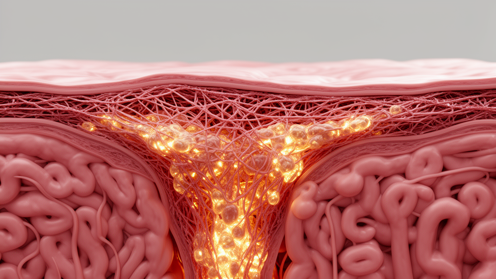
Endometriosis and adenomyosis affect an estimated one in ten women of reproductive age, causing chronic pelvic pain, heavy menstrual bleeding, and infertility, yet their underlying biology remains stubbornly incomplete. A new narrative review published in Reproductive Sciences by Hiroshi Kobayashi of Nara Medical University argues that the field has been missing a crucial dimension: the physical mechanics of the lesions themselves. The review, titled “Endometriosis and Adenomyosis: Mechanotransduction, Microenvironment, and Mechanisms Beyond Lesions,” proposes that tissue stiffness and the cellular machinery that senses it may help explain why these diseases are so heterogeneous, why some lesions invade aggressively while others remain indolent, and why current anti-inflammatory and hormonal therapies leave so many patients undertreated.
The central premise rests on a concept called mechanotransduction, the process by which cells convert mechanical cues from their surroundings into biochemical signals. It has been known since landmark experiments published in Science in 2005 that cells can feel and respond to the stiffness of the substrate on which they grow, and subsequent work showed that matrix elasticity can even direct stem cell lineage specification. When tissue becomes stiffer, as it does in fibrosis, cells anchored to the extracellular matrix through integrin receptors experience altered tension. This tension propagates through focal adhesions and the actin cytoskeleton, activating signaling cascades involving Rho-associated kinases and the mechanosensitive transcriptional co-activators YAP and TAZ, which translocate to the nucleus and reprogram gene expression toward proliferation, invasion, and matrix production.
Kobayashi’s review assembles evidence that this mechanobiological circuitry is active in ectopic endometrial tissue. Endometriotic stromal cells have been shown to contract collagen gels more vigorously than normal endometrial stromal cells, a possible mechanism underlying endometriosis-associated fibrosis. Smooth muscle cells and myofibroblasts are frequent components of endometriotic lesions, and their distribution of smooth muscle actin and collagen differs from the surrounding peritoneal microenvironment. In adenomyosis, where endometrial tissue invades the muscular wall of the uterus, the junctional zone smooth muscle cells display abnormal activation of the RhoA/ROCK signaling pathway, and estrogen has been reported to hyperactivate this pathway, driving overproliferation of these cells. Hippo pathway inactivation, which liberates YAP to promote cell proliferation and block apoptosis, has been implicated in adenomyosis development, while activated Hippo/YAP signaling promotes proliferation and survival of endometrial stromal cells in endometriosis.
What makes the mechanical perspective potentially transformative is that it reframes the disease as a feedback loop rather than a one-way cascade. Chronic inflammation, recurrent bleeding into lesions, and progressive accumulation of extracellular matrix all contribute to tissue stiffening. That stiffening, in turn, may push cells toward greater proliferation, invasion, and further matrix deposition through integrin signaling, TGF-beta-driven myofibroblast transformation, and YAP/TAZ activation. The review highlights the reciprocal interaction: biological processes create mechanical changes, and mechanical changes amplify biological processes. This self-reinforcing cycle could explain the progressive nature of ovarian endometriomas documented in longitudinal studies and the extensive fibrosis seen in deep infiltrating disease and adenomyosis, where fibrosis extent correlates with heavy menstrual bleeding.
Importantly, the review is candid about the limits of the evidence. Direct measurements of tissue stiffness in these diseases do exist. Transvaginal elastosonography and shear wave elastography have been used to diagnose deep endometriosis and adenomyosis, shear wave elastography values have been characterized in endometriomas with machine learning prediction models, and magnetic resonance elastography has shown feasibility for diagnosing uterine adenomyosis. A systematic review with meta-analysis found ultrasound elastography promising for diagnosing both conditions. However, standardized comparisons of stiffness across different lesion types remain limited, and much of the mechanotransduction evidence derives from in vitro experiments, animal models, or extrapolation from other fibrotic diseases such as liver fibrosis, pulmonary fibrosis, keloids, and atherosclerosis. Direct causal relationships between stiffness and lesion behavior in human endometriosis and adenomyosis have not been firmly established.
The mechanobiological framework does not replace established concepts but complements them. The review weaves tissue mechanics together with the classical pillars of endometriosis biology: estrogen signaling and local aromatase activity, inflammatory cytokines in peritoneal fluid, immune dysregulation including macrophage infiltration, oxidative stress from iron released by repeated hemorrhage, angiogenesis, and neurogenesis. Each of these intersects with mechanics. TGF-beta signaling, a master driver of fibrosis, coincides with epithelial-mesenchymal transition and fibroblast-to-myofibroblast transdifferentiation in adenomyosis, and macrophage-to-myofibroblast transitions driven by TGFB1/SMAD3 signaling promote fibrosis progression in endometriosis. Meanwhile, YAP-mediated mechanotransduction has been shown to tune the inflammatory response of macrophages, linking stiffness directly to the immune microenvironment. Reactive oxygen species and TGF-beta regulate each other in what researchers have called a perverse cycle for fibrosis, and matrix stiffness crosstalk with reactive oxygen species drives endometrial repair through the HIF-1alpha/YAP axis during menstruation.
The vascular and nervous dimensions of the lesions also acquire new meaning under a mechanical lens. Angiogenesis and lymphangiogenesis are increased in adenomyosis, visualized by multiplex immunohistochemistry, and mechanoregulation of vascular endothelial growth factor receptor 2 is known to shape angiogenesis generally. Rich innervation of deep infiltrating endometriosis and adenomyotic tissue is well documented, with nerve fiber density proposed as a biomarker, and perineural invasion contributes to endometriosis-associated pain. Stiff, fibrotic tissue may physically compress and sensitize these nerves while the biochemical milieu of inflammatory and neurogenic mediators amplifies pain signaling. Platelets, which accumulate at sites of cyclic bleeding, induce endothelial-mesenchymal and mesothelial-mesenchymal transitions that fuel fibrogenesis, and experimental work suggests anti-platelet therapy holds promise for adenomyosis, adding another candidate node in the mechanical-biological network.
Therapeutically, the review points to several mechanotransduction nodes already under investigation. The Rho kinase inhibitor fasudil has been shown to inhibit the proliferation and contractility of endometriotic stromal cells and to induce cell cycle arrest and apoptosis, making it a candidate agent for endometriosis treatment. Focal adhesion kinase-mediated sequences covering cell adhesion, inflammatory response, and fibrosis have been proposed as therapeutic targets. Targeting hypoxia-mediated YAP1 nuclear translocation ameliorated endometriosis pathogenesis in experimental models without compromising maternal fertility, and the SOX18-OTUB1-YAP1 axis has been nominated as a new endometriosis target. If tissue stiffness proves to be a driver rather than merely a byproduct of disease, elastography could evolve from a diagnostic tool into a longitudinal biomarker tracking lesion progression and treatment response, and mechanical microenvironment profiling could guide lesion-specific therapy.
The review’s most consequential proposal may be its framing of lesion heterogeneity. Endometriosis and adenomyosis are increasingly viewed as a spectrum with shared pathophysiology, yet superficial peritoneal lesions, ovarian endometriomas, deep infiltrating nodules, and adenomyotic foci behave very differently. Kobayashi argues that lesion phenotypes reflect interactions among lesion origin, cell-intrinsic properties such as genetic and epigenetic alterations, and the local microenvironment, of which mechanical properties are an underappreciated component. Spatial transcriptomics has already revealed distinct penetration features along the path from invaginating site to deep lesion in adenomyosis, and single-cell profiling has identified distinct hormonal, immunologic, and inflammatory signatures among endometriosis-constituting cells. Adding mechanical parameters to such lesion-level maps could finally explain why identical cells behave differently in different niches.
What is needed next, the review concludes, are longitudinal, lesion-level studies that integrate mechanical and biological measurements in the same patients over time. Such studies would test whether stiffness precedes and predicts fibrosis progression, whether mechanotransduction pathway activation correlates with clinical outcomes like pain and infertility, and whether mechanical biomarkers can stratify patients for existing or emerging therapies. Until then, the mechanobiology of endometriosis and adenomyosis remains a hypothesis-generating framework rather than settled science, but one that offers a fresh conceptual vocabulary for a disease that has long been described almost exclusively in chemical and cellular terms. For millions of patients awaiting better answers, the idea that their lesions may be governed partly by physics, and that physics can be measured, modeled, and perhaps manipulated, is a provocative and hopeful shift in perspective.
Subject of Research: Mechanotransduction and tissue stiffness in the pathogenesis of endometriosis and adenomyosis
Article Title: Endometriosis and Adenomyosis: Mechanotransduction, Microenvironment, and Mechanisms Beyond Lesions
Article References: Kobayashi, H. (2026). Endometriosis and Adenomyosis: Mechanotransduction, Microenvironment, and Mechanisms Beyond Lesions. Reproductive Sciences. https://doi.org/10.1007/s43032-026-02237-x
Image Credits: AI Generated
DOI: 10.1007/s43032-026-02237-x
Keywords: endometriosis, adenomyosis, mechanotransduction, tissue stiffness, fibrosis, YAP/TAZ, TGF-beta, elastography, microenvironment, inflammation, integrin signaling, reproductive health
Cite Scienmag News
Ophelia Keating. (October 9, 2026). Stiff Tissue, Sick Tissue: How Mechanics May Drive Endometriosis and Adenomyosis. Scienmag. https://scienmag.com/stiff-tissue-sick-tissue-how-mechanics-may-drive-endometriosis-and-adenomyosis/
Ophelia Keating. "Stiff Tissue, Sick Tissue: How Mechanics May Drive Endometriosis and Adenomyosis." Scienmag, 9 October 2026, https://scienmag.com/stiff-tissue-sick-tissue-how-mechanics-may-drive-endometriosis-and-adenomyosis/. Accessed 9 October 2026.
Ophelia Keating. "Stiff Tissue, Sick Tissue: How Mechanics May Drive Endometriosis and Adenomyosis." Scienmag. October 9, 2026. https://scienmag.com/stiff-tissue-sick-tissue-how-mechanics-may-drive-endometriosis-and-adenomyosis/


