


Optic neuritis, the inflammatory demyelination of the optic nerve that blurs or dims vision in one eye over days, has long been one of neurology’s most deceptively difficult diagnoses. Its clinical picture varies widely: some patients present with textbook pain on eye movement and a strikingly impaired optic disc, while others arrive with subtle symptoms that overlap with ischemic, compressive, or hereditary optic neuropathies. In 2022, an international consortium published consensus criteria, known as the ICON criteria, to standardize how clinicians classify the condition. Yet those criteria were built largely on expert opinion rather than prospective validation, and a new study from Barcelona now suggests they may be too strict for real-world practice, missing nearly half of patients who ultimately prove to have the disease.
The research, led by Luca Bollo and Angela Vidal-Jordana at the Multiple Sclerosis Centre of Catalonia in collaboration with Vall d’Hebron University Hospital and published in the Journal of Neurology, prospectively followed 46 patients experiencing their first episode of unilateral subacute optic neuritis. Each participant underwent a comprehensive workup: detailed clinical examination, orbital and brain magnetic resonance imaging, optical coherence tomography of the retinal nerve fiber layer, laboratory testing including serum antibodies against aquaporin-4 and myelin oligodendrocyte glycoprotein, and visual evoked potentials, a neurophysiological test that measures how efficiently the visual pathway conducts signals from the retina to the visual cortex. The investigators then applied the original ICON 2022 criteria and compared the results with two modified versions of the framework.
The headline finding is stark. Under the original ICON 2022 criteria, only 20 of the 46 patients, or 43.5 percent, reached the category of definite optic neuritis. When the researchers loosened the clinical requirements, allowing a diagnosis with incomplete clinical features, and required two abnormal paraclinical tests among MRI, optical coherence tomography, and laboratory findings, the proportion of definite cases rose to 32 patients, or 69.6 percent, a difference that was highly statistically significant. When visual evoked potentials were added to the paraclinical toolkit alongside the less stringent clinical definitions, the figure climbed further still, to 39 patients, or 84.8 percent, nearly double the yield of the original criteria.
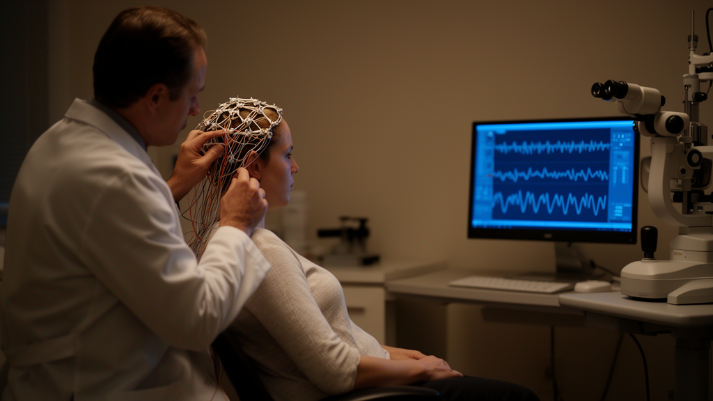
To understand why this matters, it helps to unpack what each test actually measures. Magnetic resonance imaging with orbital sequences can reveal T2-hyperintense signal and gadolinium enhancement along the optic nerve, direct evidence of inflammation and blood-nerve barrier disruption. Optical coherence tomography quantifies the thickness of the retinal nerve fiber layer and the ganglion cell complex, detecting the axonal loss that follows an inflammatory attack. Visual evoked potentials, by contrast, probe function rather than structure: a flashing or pattern-reversing checkerboard stimulus is presented to each eye while electrodes over the occipital cortex record the timing and amplitude of the brain’s response. Demyelination delays the P100 latency, the characteristic peak of the response, often before structural thinning becomes measurable on scans.
This functional sensitivity is precisely why the addition of VEPs proved so powerful in the study. Patients with incomplete clinical presentations, for example those without the classic combination of pain, visual acuity loss, color vision impairment, and a relative afferent pupillary defect, could still be classified as definite optic neuritis when their paraclinical workup was typical. The effect was most pronounced in exactly this subgroup: individuals whose symptoms did not tick every clinical box but whose MRI, OCT, and electrophysiology told an unambiguous story of optic nerve inflammation. In clinical terms, the modified criteria rescued a substantial fraction of patients who would otherwise have been left in diagnostic limbo, a category with real consequences for treatment decisions, follow-up intensity, and counseling about the risk of future demyelinating events such as multiple sclerosis.
The Barcelona group’s work does not stand in isolation. Since the ICON criteria appeared in The Lancet Neurology in 2022, several groups have tested them against independent cohorts and reported similar concerns about stringency. Correspondence in the same journal questioned how the criteria performed in atypical presentations, and the Acute Optic Neuritis Network published an application of the criteria in 2024 that highlighted classification gaps. Meanwhile, the field of demyelinating disease diagnostics has been moving rapidly toward incorporating the optic nerve more broadly. The 2024 revisions of the McDonald criteria for multiple sclerosis, the most consequential diagnostic framework in the field, now allow optic nerve involvement, demonstrated by MRI or by visual evoked potentials, to contribute to the demonstration of dissemination in space, a change that formally elevates VEPs from a niche neurophysiological tool to a first-line diagnostic instrument.
Against that backdrop, the new study offers prospective, patient-level evidence that the optic neuritis criteria should evolve in the same direction. The authors’ proposed modification is conceptually simple: rather than demanding a complete clinical syndrome, the framework would accept a less strict clinical category and then require corroboration from two abnormal paraclinical tests, with VEPs counted alongside MRI, OCT, and serology. This modular architecture mirrors how modern neurology increasingly thinks about diagnosis, as a Bayesian exercise in which each test contributes independent information about the probability of disease. A delayed P100 latency on VEPs carries information that is partly independent of a normal-appearing retinal nerve fiber layer on OCT, and both can be informative when the MRI shows only subtle optic nerve signal change.
There are important caveats. The cohort of 46 patients is modest, and all participants were recruited at specialized centers in Catalonia with access to expert neuroradiology, neuro-ophthalmology, and neurophysiology, so the modified criteria will need validation in larger and more diverse populations, including settings where antibody testing and VEP recording are less readily available. The study also focused on first-episode unilateral optic neuritis, leaving open the question of how the modified framework performs in bilateral, recurrent, or pediatric disease, where differential diagnoses such as neuromyelitis optica spectrum disorder and MOG antibody-associated disease loom larger and where serological markers carry decisive weight. The researchers themselves frame their findings as supporting revisions aimed at broader clinical applicability rather than as a finished replacement for the 2022 consensus.
Even so, the implications for patients are tangible. A person who wakes with a sore, blurry eye but whose examination findings are incomplete currently risks being labeled with uncertain optic neuropathy, delaying disease-modifying therapy if multiple sclerosis is the underlying cause, or delaying plasma exchange and immunosuppression if aquaporin-4 antibody disease is suspected. By formally recognizing that paraclinical evidence, especially electrophysiological evidence from visual evoked potentials, can compensate for an atypical clinical picture, the modified criteria promise faster, more confident classification and, downstream, better-targeted treatment. The study was funded by the Instituto de Salud Carlos III, and its datasets are available from the corresponding authors, an invitation for other centers to stress-test the approach.
The broader lesson reaches beyond one disease. Diagnostic criteria in neurology have historically leaned heavily on clinical acumen, with imaging and laboratory tests playing supporting roles. The trajectory now visible across multiple sclerosis, neuromyelitis optica, MOG antibody disease, and optic neuritis points toward criteria that fuse clinical features with structural imaging, retinal layer quantification, serology, and neurophysiology in a single probabilistic framework. The Barcelona study demonstrates concretely what that fusion can achieve: the proportion of patients confidently classified as having definite optic neuritis nearly doubled, from 43.5 to 84.8 percent, simply by trusting the objective signals that modern technology can already deliver. If subsequent multicenter validation confirms these results, the humble visual evoked potential, a test first described decades ago, may finally take its place at the center of optic nerve diagnostics, and fewer patients will be left waiting in the gray zone between suspicion and certainty.
Subject of Research: Prospective evaluation of the 2022 international consensus criteria for optic neuritis and the diagnostic value of visual evoked potentials
Article Title: Evaluation of the 2022 international consensus criteria for optic neuritis: the role of clinical features and visual evoked potentials
Article References: Bollo, L., Vidal-Jordana, A., Rodríguez-Acevedo, B., Sceppacuercia, S., Mongay-Ochoa, N., Zancan, V., Ajdinaj, P., Braga, N., Cabello, S., Alberich, M., Corral, J., Auger, C., Tintoré, M., Montalban, X., Pareto, D., Moncho, D., Rovira, À., & Sastre-Garriga, J. (2026). Evaluation of the 2022 international consensus criteria for optic neuritis: the role of clinical features and visual evoked potentials. Journal of Neurology, 273(10), Article 561. https://doi.org/10.1007/s00415-026-13972-1
Image Credits: AI Generated
DOI: 10.1007/s00415-026-13972-1
Keywords: optic neuritis, diagnostic criteria, visual evoked potentials, ICON criteria, MRI, optical coherence tomography, multiple sclerosis, demyelination, neurology, optic nerve, MOG antibodies, prospective study
Cite Scienmag News
Ophelia Keating. (October 9, 2026). Adding Visual Evoked Potentials to 2022 Optic Neuritis Criteria Nearly Doubles Definite Diagnoses. Scienmag. https://scienmag.com/adding-visual-evoked-potentials-to-2022-optic-neuritis-criteria-nearly-doubles-definite-diagnoses/
Ophelia Keating. "Adding Visual Evoked Potentials to 2022 Optic Neuritis Criteria Nearly Doubles Definite Diagnoses." Scienmag, 9 October 2026, https://scienmag.com/adding-visual-evoked-potentials-to-2022-optic-neuritis-criteria-nearly-doubles-definite-diagnoses/. Accessed 9 October 2026.
Ophelia Keating. "Adding Visual Evoked Potentials to 2022 Optic Neuritis Criteria Nearly Doubles Definite Diagnoses." Scienmag. October 9, 2026. https://scienmag.com/adding-visual-evoked-potentials-to-2022-optic-neuritis-criteria-nearly-doubles-definite-diagnoses/


